Faculty of Dental Medicine, Institute of Dental Sciences, Hebrew University, Jerusalem, Israel.
Orthopaedic Department, Hadassah - Hebrew University Medical Center, Jerusalem, Israel.
J Cell Mol Med. 2016 May;20(5):815-24. doi: 10.1111/jcmm.12762. Epub 2016 Feb 24.
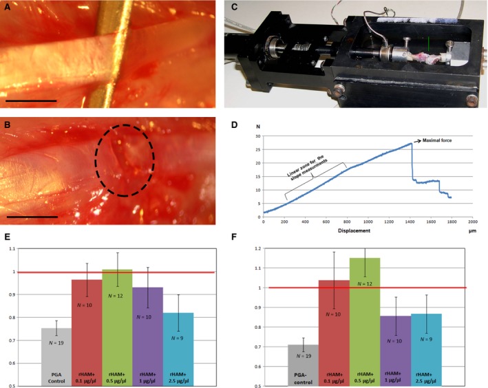
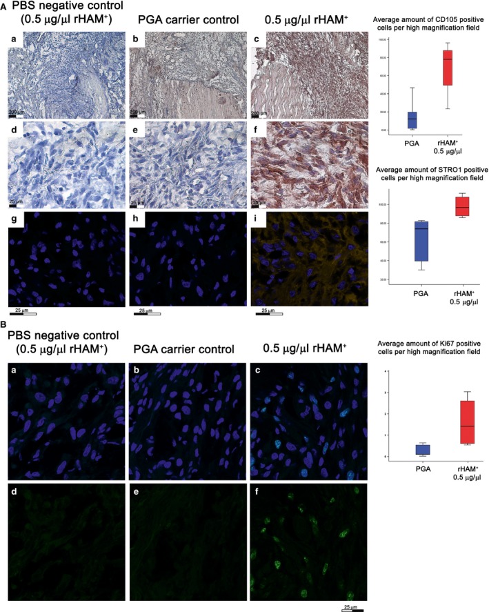
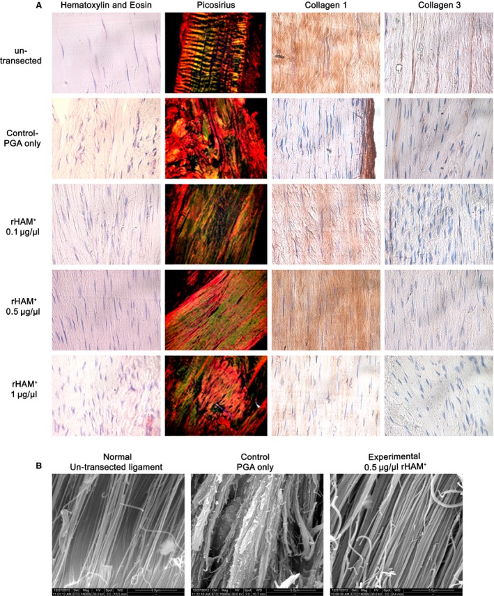
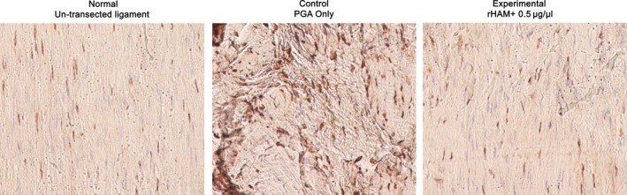
Injuries to ligaments are common, painful and debilitating, causing joint instability and impaired protective proprioception sensation around the joint. Healing of torn ligaments usually fails to take place, and surgical replacement or reconstruction is required. Previously, we showed that in vivo application of the recombinant human amelogenin protein (rHAM(+)) resulted in enhanced healing of the tooth-supporting tissues. The aim of this study was to evaluate whether amelogenin might also enhance repair of skeletal ligaments. The rat knee medial collateral ligament (MCL) was chosen to prove the concept. Full thickness tear was created and various concentrations of rHAM(+), dissolved in propylene glycol alginate (PGA) carrier, were applied to the transected MCL. 12 weeks after transection, the mechanical properties, structure and composition of transected ligaments treated with 0.5 μg/μl rHAM(+) were similar to the normal un-transected ligaments, and were much stronger, stiffer and organized than control ligaments, treated with PGA only. Furthermore, the proprioceptive free nerve endings, in the 0.5 μg/μl rHAM(+) treated group, were parallel to the collagen fibres similar to their arrangement in normal ligament, while in the control ligaments the free nerve endings were entrapped in the scar tissue at different directions, not parallel to the axis of the force. Four days after transection, treatment with 0.5 μg/μl rHAM(+) increased the amount of cells expressing mesenchymal stem cell markers at the injured site. In conclusion application of rHAM(+) dose dependently induced mechanical, structural and sensory healing of torn skeletal ligament. Initially the process involved recruitment and proliferation of cells expressing mesenchymal stem cell markers.
韧带损伤很常见,疼痛且使人虚弱,导致关节不稳定,并损害关节周围的保护性本体感觉。撕裂的韧带通常无法愈合,需要进行手术替代或重建。此前,我们已经表明,重组人釉原蛋白(rHAM(+))的体内应用可增强对牙齿支持组织的愈合作用。本研究旨在评估釉原蛋白是否也能增强对骨骼韧带的修复作用。我们选择大鼠膝关节内侧副韧带(MCL)来验证这一概念。在 MCL 上造成全层撕裂,然后将不同浓度的 rHAM(+)溶解在藻酸丙二醇酯(PGA)载体中,应用于横断的 MCL。横断后 12 周,用 0.5 μg/μl rHAM(+)处理的横断韧带的机械性能、结构和组成与正常未横断的韧带相似,且比仅用 PGA 处理的对照组韧带更强、更硬且更有组织。此外,在 0.5 μg/μl rHAM(+)处理组中,本体感觉游离神经末梢与胶原纤维平行排列,类似于正常韧带中的排列方式,而在对照组韧带中,游离神经末梢被包裹在不同方向的瘢痕组织中,与力的轴不平行。横断后 4 天,用 0.5 μg/μl rHAM(+)处理可增加受伤部位表达间充质干细胞标志物的细胞数量。总之,rHAM(+)的应用剂量依赖性地诱导了撕裂的骨骼韧带的机械、结构和感觉愈合。最初,这个过程涉及到表达间充质干细胞标志物的细胞的募集和增殖。