Puranik Ameya D, Singh Natasha, Maheshwari Shailendra, Gupta Nitin, Wali Pravin
Department of Nuclear Medicine and PET Imaging, P.D. Hinduja National Hospital and Medical Research Center, Mumbai, Maharashtra, India.
Department of Radiology, P.D. Hinduja National Hospital and Medical Research Center, Mumbai, Maharashtra, India.
Indian J Nucl Med. 2016 Jan-Mar;31(1):39-41. doi: 10.4103/0972-3919.172357.
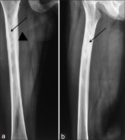
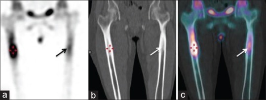
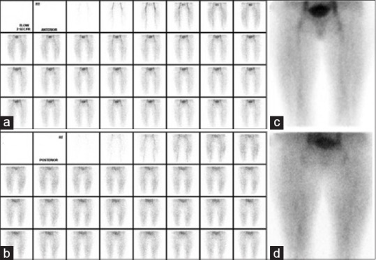
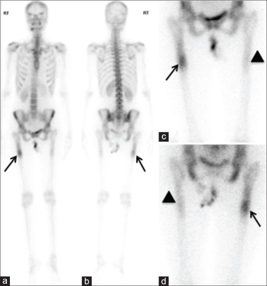
Sclerotic bone dysplasias are diagnosed based on clinical and radiological features; however, in some instances pose a dilemma. We herewith report a case of a 38-year-old female who presented with right lower extremity pain, and was detected to have sclerotic diaphyseal lesion on X-ray. Triphasic 99mTc methylene diphosphonate (MDP) Bone scan helped in confirming the diagnosis of intramedullary osteosclerosis, a dysplastic bone disorder.
骨硬化发育异常是根据临床和放射学特征进行诊断的;然而,在某些情况下会造成诊断困难。我们在此报告一例38岁女性,她因右下肢疼痛就诊,X线检查发现骨干硬化性病变。三相99mTc亚甲基二膦酸盐(MDP)骨扫描有助于确诊为骨髓性骨硬化,这是一种发育异常性骨病。