• 文献检索
  • 文档翻译
  • 深度研究
  • 学术资讯
  • Suppr Zotero 插件Zotero 插件
  • 邀请有礼
  • 套餐&价格
  • 历史记录
应用&插件
Suppr Zotero 插件Zotero 插件浏览器插件Mac 客户端Windows 客户端微信小程序
定价
高级版会员购买积分包购买API积分包
服务
文献检索文档翻译深度研究API 文档MCP 服务
关于我们
关于 Suppr公司介绍联系我们用户协议隐私条款
关注我们

Suppr 超能文献

核心技术专利:CN118964589B侵权必究
粤ICP备2023148730 号-1Suppr @ 2026

文献检索

告别复杂PubMed语法,用中文像聊天一样搜索,搜遍4000万医学文献。AI智能推荐,让科研检索更轻松。

立即免费搜索

文件翻译

保留排版,准确专业,支持PDF/Word/PPT等文件格式,支持 12+语言互译。

免费翻译文档

深度研究

AI帮你快速写综述,25分钟生成高质量综述,智能提取关键信息,辅助科研写作。

立即免费体验

延髓和肢体起病的早期肌萎缩侧索硬化症的高级磁共振神经成像

Advanced magnetic resonance neuroimaging in bulbar and limb onset early amyotrophic lateral sclerosis.

作者信息

Vora Maulik, Kumar Suresh, Sharma Sanjiv, Sharma Sudhir, Makhaik Sushma, Sood R G

机构信息

Department of Radiodiagnosis and Imaging, Indira Gandhi Medical College, Shimla, Himachal Pradesh, India.

Department of Neurology, Indira Gandhi Medical College, Shimla, Himachal Pradesh, India.

出版信息

J Neurosci Rural Pract. 2016 Jan-Mar;7(1):102-8. doi: 10.4103/0976-3147.165423.

DOI:10.4103/0976-3147.165423
PMID:26933355
原文链接:https://pmc.ncbi.nlm.nih.gov/articles/PMC4750305/
Abstract

INTRODUCTION

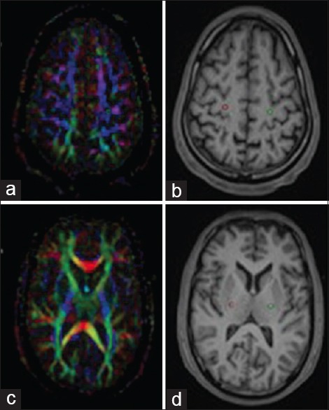
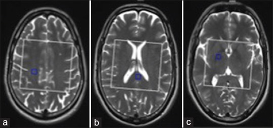
Amyotrophic lateral sclerosis (ALS) is a fatal and most common motor neuron disease, caused by progressive loss of motor neurons. Diffusion tensor imaging (DTI) and magnetic resonance spectroscopic (MRS) studies detect pathological changes in neuronal fibers in vivo. We evaluated the role of DTI and MRS in early course of the disease, which may prove beneficial in the early diagnosis and better management.

MATERIALS AND METHODS

Twenty-one patients with ALS and 13 age-matched controls received 1.5T DTI and three-dimensional multi-voxel MRS. Fractional anisotropy (FA), apparent diffusion coefficient, N-acetyl aspartate (NAA)/Creatine (Cr), and NAA/Choline (Ch) ratios were analyzed in various regions of the brain and compared with healthy controls. ALS patients were classified as definite, possible, and probable category, and patients were also studied in limb versus bulbar onset.

RESULTS

Decreased FA and increase mean diffusivity values in regions of corticospinal tract (CST) and corpus callosum (CC) was consistent finding in definite and probable disease category (P < 0.05). In possible disease, CC involvement was not significant. NAA/Cr and NAA/Ch ratios were lower in CC and regions of CST. However, in possible disease, CC involvement was not significant, while regions of CST were showing significant reduction in NAA/Cr and NAA/Ch ratios (P < 0.05).

CONCLUSION

DTI and MRS detect changes associated with ALS even in the early phase of the disease. Bulbar onset and limb onset ALS patients show different pattern of involvement. Extramotor involvement suggested by CC involvement is a feature seen in bulbar onset patient and can suggest poor outcome in such patients. The present findings may be helpful for designing further studies in the direction of more early diagnosis of disease and its management.

摘要

引言

肌萎缩侧索硬化症(ALS)是一种致命且最常见的运动神经元疾病,由运动神经元的进行性丧失引起。扩散张量成像(DTI)和磁共振波谱(MRS)研究可在体内检测神经元纤维的病理变化。我们评估了DTI和MRS在该疾病早期病程中的作用,这可能对早期诊断和更好的治疗有益。

材料与方法

21例ALS患者和13例年龄匹配的对照者接受了1.5T DTI和三维多体素MRS检查。分析了大脑各个区域的分数各向异性(FA)、表观扩散系数、N-乙酰天门冬氨酸(NAA)/肌酸(Cr)以及NAA/胆碱(Ch)比值,并与健康对照者进行比较。ALS患者被分为确诊、可能和疑似类别,还对肢体起病与延髓起病的患者进行了研究。

结果

在确诊和疑似疾病类别中,皮质脊髓束(CST)和胼胝体(CC)区域的FA降低和平均扩散率值增加是一致的发现(P < 0.05)。在可能的疾病中,CC受累不显著。CC和CST区域的NAA/Cr和NAA/Ch比值较低。然而,在可能的疾病中,CC受累不显著,而CST区域的NAA/Cr和NAA/Ch比值显著降低(P < 0.05)。

结论

DTI和MRS即使在疾病早期也能检测到与ALS相关的变化。延髓起病和肢体起病的ALS患者表现出不同的受累模式。CC受累提示的运动外受累是延髓起病患者的一个特征,可提示此类患者预后不良。目前的发现可能有助于在疾病更早期诊断及其治疗方向上设计进一步的研究。

https://cdn.ncbi.nlm.nih.gov/pmc/blobs/3cc4/4750305/00a2ec1f0360/JNRP-7-102-g002.jpg
https://cdn.ncbi.nlm.nih.gov/pmc/blobs/3cc4/4750305/205b8fecfd33/JNRP-7-102-g001.jpg
https://cdn.ncbi.nlm.nih.gov/pmc/blobs/3cc4/4750305/00a2ec1f0360/JNRP-7-102-g002.jpg
https://cdn.ncbi.nlm.nih.gov/pmc/blobs/3cc4/4750305/205b8fecfd33/JNRP-7-102-g001.jpg
https://cdn.ncbi.nlm.nih.gov/pmc/blobs/3cc4/4750305/00a2ec1f0360/JNRP-7-102-g002.jpg

相似文献

1
Advanced magnetic resonance neuroimaging in bulbar and limb onset early amyotrophic lateral sclerosis.延髓和肢体起病的早期肌萎缩侧索硬化症的高级磁共振神经成像
J Neurosci Rural Pract. 2016 Jan-Mar;7(1):102-8. doi: 10.4103/0976-3147.165423.
2
Role of diffusion tensor imaging or magnetic resonance spectroscopy in the diagnosis and disability assessment of amyotrophic lateral sclerosis.扩散张量成像或磁共振波谱在肌萎缩侧索硬化症诊断及功能障碍评估中的作用
J Neurol Sci. 2015 Jan 15;348(1-2):206-10. doi: 10.1016/j.jns.2014.12.004. Epub 2014 Dec 6.
3
Differential involvement of corticospinal tract (CST) fibers in UMN-predominant ALS patients with or without CST hyperintensity: A diffusion tensor tractography study.皮质脊髓束(CST)纤维在伴有或不伴有CST高信号的以UMN为主的肌萎缩侧索硬化症患者中的差异参与:一项扩散张量纤维束成像研究。
Neuroimage Clin. 2017 Feb 22;14:574-579. doi: 10.1016/j.nicl.2017.02.017. eCollection 2017.
4
Integrated imaging of [C]-PBR28 PET, MR diffusion and magnetic resonance spectroscopy H-MRS in amyotrophic lateral sclerosis.肌萎缩侧索硬化症的 [C]-PBR28 PET、MR 扩散和磁共振波谱 H-MRS 联合成像。
Neuroimage Clin. 2018 Aug 9;20:357-364. doi: 10.1016/j.nicl.2018.08.007. eCollection 2018.
5
Diffusion tensor imaging patterns differ in bulbar and limb onset amyotrophic lateral sclerosis.延髓起病和肢体起病的肌萎缩侧索硬化症的弥散张量成像模式存在差异。
Clin Neurol Neurosurg. 2013 Aug;115(8):1281-7. doi: 10.1016/j.clineuro.2012.11.031. Epub 2012 Dec 23.
6
Diffusion tensor MRI and MR spectroscopy in long lasting upper motor neuron involvement in amyotrophic lateral sclerosis.扩散张量磁共振成像和磁共振波谱在肌萎缩侧索硬化症长期上运动神经元受累中的应用
Arch Ital Biol. 2009 Sep;147(3):69-82.
7
Combined MR spectroscopic imaging and diffusion tensor MRI visualizes corticospinal tract degeneration in amyotrophic lateral sclerosis.联合磁共振波谱成像和扩散张量磁共振成像可显示肌萎缩侧索硬化症中的皮质脊髓束变性。
J Neurol. 2004 Oct;251(10):1249-54. doi: 10.1007/s00415-004-0526-9.
8
Amyotrophic lateral sclerosis: diffusion-tensor and chemical shift MR imaging at 3.0 T.肌萎缩侧索硬化症:3.0 T磁共振扩散张量成像和化学位移成像
Radiology. 2006 Jun;239(3):831-8. doi: 10.1148/radiol.2393050573. Epub 2006 Apr 26.
9
A proton magnetic resonance spectroscopic study in ALS: correlation with clinical findings.肌萎缩侧索硬化症的质子磁共振波谱研究:与临床发现的相关性
Neurology. 1998 Oct;51(4):1104-9. doi: 10.1212/wnl.51.4.1104.
10
Interhemispheric connectivity in amyotrophic lateral sclerosis: A near-infrared spectroscopy and diffusion tensor imaging study.肌萎缩侧索硬化症的半球间连接性:一项近红外光谱和扩散张量成像研究。
Neuroimage Clin. 2016 Sep 29;12:666-672. doi: 10.1016/j.nicl.2016.09.020. eCollection 2016.

引用本文的文献

1
Premotor cortex and frontal eye field region metabolite alteration in human amyotrophic lateral sclerosis patients: A quantitative survey.肌萎缩侧索硬化症患者运动前皮质和额叶眼动区代谢物变化:一项定量研究。
Neuroradiol J. 2025 May 21:19714009251345102. doi: 10.1177/19714009251345102.
2
Hippocampal Metabolic Alterations in Amyotrophic Lateral Sclerosis: A Magnetic Resonance Spectroscopy Study.肌萎缩侧索硬化症中海马体的代谢改变:一项磁共振波谱研究
Life (Basel). 2023 Feb 17;13(2):571. doi: 10.3390/life13020571.
3
Magnetic Resonance Spectroscopy in the Ventral Tegmental Area Distinguishes Responders to Suvorexant Prior to Treatment: A 4-Week Prospective Cohort Study.

本文引用的文献

1
Central white matter degeneration in bulbar- and limb-onset amyotrophic lateral sclerosis.延髓和肢体起病的肌萎缩侧索硬化症中的中枢白质变性
J Neurol. 2014 Oct;261(10):1961-7. doi: 10.1007/s00415-014-7434-4. Epub 2014 Jul 25.
2
25 years of neuroimaging in amyotrophic lateral sclerosis.25 年的肌萎缩侧索硬化症神经影像学研究。
Nat Rev Neurol. 2013 Sep;9(9):513-24. doi: 10.1038/nrneurol.2013.153. Epub 2013 Aug 6.
3
Diffusion tensor MRI of the corpus callosum in amyotrophic lateral sclerosis.肌萎缩侧索硬化症患者胼胝体的扩散张量磁共振成像
腹侧被盖区的磁共振波谱在治疗前可区分对苏沃雷生有反应者:一项为期4周的前瞻性队列研究。
Front Psychiatry. 2021 Aug 23;12:714376. doi: 10.3389/fpsyt.2021.714376. eCollection 2021.
4
Progressive Neurochemical Abnormalities in Cognitive and Motor Subgroups of Amyotrophic Lateral Sclerosis: A Prospective Multicenter Study.肌萎缩侧索硬化症认知和运动亚组的进行性神经化学异常:一项前瞻性多中心研究。
Neurology. 2021 Aug 24;97(8):e803-e813. doi: 10.1212/WNL.0000000000012367. Epub 2021 Jun 14.
5
Early white matter changes on diffusion tensor imaging in amyotrophic lateral sclerosis.肌萎缩侧索硬化症患者弥散张量成像早期白质改变
Ann Clin Transl Neurol. 2020 Jul;7(7):1265. doi: 10.1002/acn3.51108. Epub 2020 Jul 8.
6
Cerebral degeneration in amyotrophic lateral sclerosis: A prospective multicenter magnetic resonance spectroscopy study.肌萎缩侧索硬化症中的脑变性:一项前瞻性多中心磁共振波谱研究。
Neurol Clin Pract. 2019 Oct;9(5):400-407. doi: 10.1212/CPJ.0000000000000674.
7
Magnetic Resonance Spectroscopy in ALS.肌萎缩侧索硬化症中的磁共振波谱分析
Front Neurol. 2019 May 10;10:482. doi: 10.3389/fneur.2019.00482. eCollection 2019.
8
Clinical Measures of Bulbar Dysfunction in ALS.肌萎缩侧索硬化症中延髓功能障碍的临床测量
Front Neurol. 2019 Feb 19;10:106. doi: 10.3389/fneur.2019.00106. eCollection 2019.
J Magn Reson Imaging. 2014 Mar;39(3):641-7. doi: 10.1002/jmri.24218. Epub 2013 Jul 10.
4
Diffusion tensor imaging patterns differ in bulbar and limb onset amyotrophic lateral sclerosis.延髓起病和肢体起病的肌萎缩侧索硬化症的弥散张量成像模式存在差异。
Clin Neurol Neurosurg. 2013 Aug;115(8):1281-7. doi: 10.1016/j.clineuro.2012.11.031. Epub 2012 Dec 23.
5
Longitudinal diffusion tensor imaging in amyotrophic lateral sclerosis.肌萎缩侧索硬化症的纵向扩散张量成像。
BMC Neurosci. 2012 Nov 8;13:141. doi: 10.1186/1471-2202-13-141.
6
Repeatability and variation of region-of-interest methods using quantitative diffusion tensor MR imaging of the brain.使用脑定量扩散张量磁共振成像的感兴趣区方法的可重复性和可变性。
BMC Med Imaging. 2012 Oct 11;12:30. doi: 10.1186/1471-2342-12-30.
7
Corpus callosum area in amyotrophic lateral sclerosis.肌萎缩侧索硬化症中的胼胝体区域。
Amyotroph Lateral Scler. 2012 Oct;13(6):589-91. doi: 10.3109/17482968.2012.708935. Epub 2012 Aug 8.
8
White matter alterations differ in primary lateral sclerosis and amyotrophic lateral sclerosis.白质改变在原发性侧索硬化症和肌萎缩性侧索硬化症中有所不同。
Brain. 2011 Sep;134(Pt 9):2642-55. doi: 10.1093/brain/awr178. Epub 2011 Jul 28.
9
NERVE FIBRE DEGENERATION IN THE BRAIN IN AMYOTROPHIC LATERAL SCLEROSIS.肌萎缩侧索硬化症患者大脑中的神经纤维变性
J Neurol Neurosurg Psychiatry. 1960 Nov;23(4):269-82. doi: 10.1136/jnnp.23.4.269.
10
1H MRS of basal ganglia and thalamus in amyotrophic lateral sclerosis.1H MRS 于肌萎缩侧索硬化症基底节和丘脑的应用。
NMR Biomed. 2011 Dec;24(10):1270-6. doi: 10.1002/nbm.1687. Epub 2011 Mar 15.