Perandini Simone, Soardi Gian Alberto, Motton Massimiliano, Augelli Raffaele, Zantedeschi Lisa, Montemezzi Stefania
Department of Radiology, Azienda Ospedaliera Universitaria Integrata (AOUI) Verona, Verona, Italy.
Pol J Radiol. 2016 Feb 11;81:46-50. doi: 10.12659/PJR.895307. eCollection 2016.
A disappearing or persistent solid pulmonary nodule is a neglected clinical entity that still poses serious interpretative issues to date. Traditional knowledge deriving from previous reports suggests particular features, such as smooth edges or regular shape, to be significantly associated with benignity. A large number of benign nodules are reported among smokers in lung cancer screening programmes. The aim of this single-center retrospective study was to correlate specific imaging features to verify if traditional knowledge as well as more recent acquisitions regarding benign SPNs can be considered reliable in a current case series of nodules collected in a non-smoker cohort of patients.
MATERIAL/METHODS: Fifty-three solid SPNs proven as non-growing during follow-up imaging were analyzed with regard to their imaging features at thin-section CT, their predicted malignancy risk according to three major risk assessment models, minimum density analysis and contrast enhanced-CT in the relative subgroups of nodules which underwent such tests.
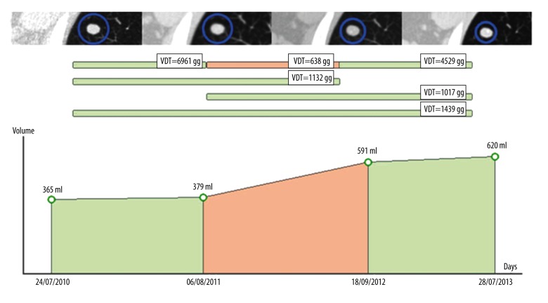
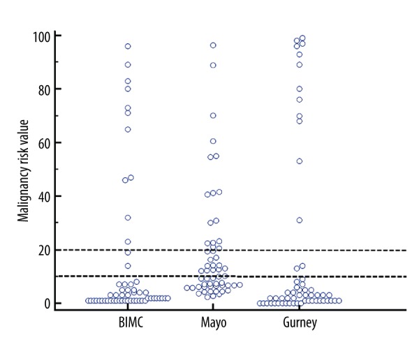
Eleven nodules disappeared during follow-up, 29 showed volume loss and 16 had a VDT of 1121 days or higher. There were 48 nodules located peripherally (85.71%). Evaluation of the enhancement after contrast media (n=29) showed mean enhancement ±SD of 25.72±35.03 HU, median of 18 HU, ranging from 0 to 190 HU. Minimum density assessment (n=30) showed mean minimum HU ±SD of -28.27±47.86 HU, median of -25 HU, ranging from -144 to 68 HU. Mean malignancy risk ±SD was 15.05±26.69% for the BIMC model, 17.22±19.00% for the Mayo Clinic model and 19.07±33.16% for the Gurney's model.
Our analysis suggests caution in using traditional knowledge when dealing with current small solid peripheral indeterminate SPNs and highlights how quantitative growth at follow-up should be the cornerstone of characterization.
消失性或持续性实性肺结节是一个被忽视的临床实体,至今仍存在严重的解读问题。以往报告中的传统认知表明,某些特征,如边缘光滑或形状规则,与良性显著相关。在肺癌筛查项目中,吸烟者中报告了大量良性结节。本单中心回顾性研究的目的是关联特定的影像学特征,以验证在当前一组非吸烟患者队列中收集的结节病例系列中,关于良性实性肺结节的传统认知以及最新研究结果是否可靠。
材料/方法:对53个在随访影像学检查中被证实无生长的实性肺结节进行分析,包括其在薄层CT上的影像学特征、根据三种主要风险评估模型预测的恶性风险、最小密度分析以及在接受此类检查的结节相对亚组中的对比增强CT。
11个结节在随访期间消失,29个显示体积缩小,16个的体积倍增时间为1121天或更长。48个结节位于周边(85.71%)。对比剂增强评估(n = 29)显示平均增强±标准差为25.72±35.03 HU,中位数为18 HU,范围为0至190 HU。最小密度评估(n = 30)显示平均最小HU±标准差为-28.27±47.86 HU,中位数为-25 HU,范围为-144至68 HU。BIMC模型的平均恶性风险±标准差为15.05±26.69%,梅奥诊所模型为17.22±19.00%,格尼模型为19.07±33.16%。
我们的分析表明,在处理当前的小型实性周边不确定肺结节时,使用传统认知需谨慎,并强调随访时的定量生长应是特征描述的基石。