Perry Chava, Lerman Hedva, Joffe Erel, Sarid Nadav, Amit Odelia, Avivi Irit, Kesler Mikhail, Ben-Ezra Jonathan, Even-Sapir Einat, Herishanu Yair
From the Department of Hematology (CP, EJ, NS, OA, IA, YH); Department of Nuclear Medicine (HL, MK, EES); Department of Pathology, Tel Aviv Sourasky Medical Center (B-E); and Sackler Faculty of Medicine, Tel Aviv University, Tel Aviv, Israel (B-E, EES, IA, YH).
Medicine (Baltimore). 2016 Mar;95(9):e2910. doi: 10.1097/MD.0000000000002910.
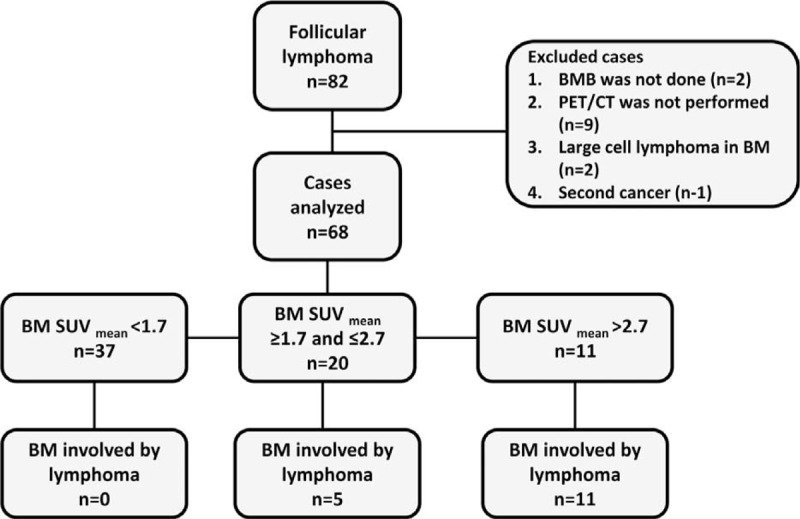
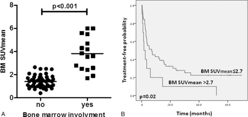
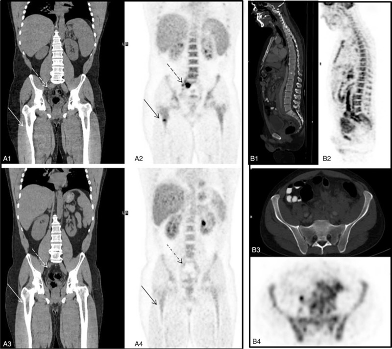
Follicular lymphoma (FL) is the 2nd most common type of lymphoma diagnosed in the Western World. Bone marrow (BM) involvement is an adverse prognostic factor in FL, routinely assessed by an arbitrary biopsy of the iliac crest. This study was aimed to investigate the role of positron emission tomography/computed tomography (PET/CT) in identifying BM involvement by FL. In this retrospective, single-center study we reviewed the records of consecutive patients with FL at diagnosis or relapse who underwent staging/restaging workup visual assessment of BM uptake was categorized as either normal, diffusely increased, or focally increased. Quantitative BM fluorine-18-fluro-deoxyglucose (FDG) uptake was measured using mean standardized uptake value (BM-SUVmean). The diagnosis of BM involvement was based on either BM histological findings or disappearance of increased uptake at end-treatment PET/CT in patients who responded to treatment. Sixty eight cases with FL were included. Sixteen (23.5%) had BM involvement, 13 (19.1%) had a biopsy proven involvement, and 3 (4.4%) had a negative BM biopsy, but increased medullary uptake that normalized post-treatment. BM FDG uptake in these patients was diffuse in 8 (50%) and focal in 8 (50%). Focal increased uptake was indicative of BM involvement; however, diffuse uptake was associated with 17 false positive cases (32.7%). Overall, visual assessment of BM involvement had a negative predictive value (NPV) of 100% and a positive predictive value (PPV) of 48.5%. On a quantitative assessment, BM-SUVmean was significantly higher in patients with BM involvement (SUVmean of 3.7 [1.7-6] vs 1.4 [0.4-2.65], P < 0.001). On receiver operator curve (ROC) analysis, BM-SUVmean > 2.7 had a PPV of 100% for BM involvement (sensitivity of 68%), while BM-SUVmean < 1.7 had an NPV of 100% (specificity of 73%). Visual assessment of PET/CT is appropriate for ruling out BM involvement by FL. Although focal increased uptake indicates marrow involvement, diffuse uptake is nonspecific. SUV measurement improves PET/CT diagnostic accuracy, identifying additional 19% of patients with BM involvement that would have been otherwise missed.
滤泡性淋巴瘤(FL)是西方世界诊断出的第二常见淋巴瘤类型。骨髓(BM)受累是FL的不良预后因素,通常通过髂嵴的任意活检进行评估。本研究旨在调查正电子发射断层扫描/计算机断层扫描(PET/CT)在识别FL骨髓受累中的作用。在这项回顾性单中心研究中,我们回顾了诊断或复发时接受分期/再分期检查的连续FL患者的记录,骨髓摄取的视觉评估分为正常、弥漫性增加或局灶性增加。使用平均标准化摄取值(BM-SUVmean)测量骨髓氟-18-氟脱氧葡萄糖(FDG)摄取量。骨髓受累的诊断基于骨髓组织学结果或治疗反应良好的患者在治疗结束时PET/CT上摄取增加的消失情况。纳入68例FL病例。16例(23.5%)有骨髓受累,13例(19.1%)经活检证实受累,3例(4.4%)骨髓活检阴性,但骨髓摄取增加,治疗后恢复正常。这些患者的骨髓FDG摄取8例(50%)为弥漫性,8例(50%)为局灶性。局灶性摄取增加提示骨髓受累;然而,弥漫性摄取与17例假阳性病例(32.7%)相关。总体而言,骨髓受累的视觉评估阴性预测值(NPV)为100%,阳性预测值(PPV)为48.5%。在定量评估中,骨髓受累患者的BM-SUVmean显著更高(SUVmean为3.7[1.7 - 6]对1.4[0.4 - 2.65],P<0.001)。在受试者操作特征曲线(ROC)分析中,BM-SUVmean>2.7对骨髓受累的PPV为100%(敏感性为68%),而BM-SUVmean<1.7的NPV为100%(特异性为73%)。PET/CT的视觉评估适用于排除FL的骨髓受累。虽然局灶性摄取增加提示骨髓受累,但弥漫性摄取不具有特异性。SUV测量提高了PET/CT的诊断准确性,可识别出另外19%原本会被漏诊的骨髓受累患者。