Cui Dan-Mo, Han De-Min, Nicolas Busaba, Hu Chang-Long, Wu Jun, Su Min-Min
Department of Otolaryngology, Head and Neck Surgery, Beijing Tongren Hospital, Capital Medical University, Beijing 100730; Key Laboratory of Otorhinolaryngology Head and Neck Surgery, Ministry of Education, Beijing Institute of Otorhinolaryngology, Beijing 100005, China.
Chin Med J (Engl). 2016 Mar 20;129(6):651-6. doi: 10.4103/0366-6999.177971.
Obstructive sleep apnea (OSA) is a common sleep disorder and is characterized by airway collapse at multiple levels of upper airway. The effectiveness of nasal surgery has been discussed in several studies and shows a promising growing interest. In this study, we intended to evaluate the effects of nasal surgery on the upper airway dimensions in patients with OSA using three-dimensional (3D) reconstruction of cone-beam computed tomography (CT).
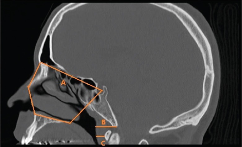
Twelve patients with moderate to severe OSA who underwent nasal surgery were included in this study. All patients were diagnosed with OSA using polysomnography (PSG) in multi sleep health centers associated with Massachusetts General Hospital, Massachusetts Eye and Ear Infirmary and the Partners Health Care from May 31, 2011 to December 14, 2013. The effect of nasal surgery was evaluated by the examination of PSG, subjective complains, and 3D reconstructed CT scan. Cross-sectional area was measured in eleven coronal levels, and nasal cavity volume was evaluated from anterior nasal spine to posterior nasal spine. The thickness of soft tissue in oral pharynx region was also measured.
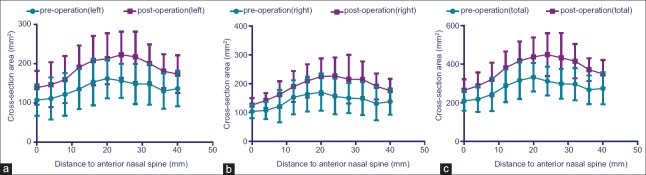
Five out of the 12 patients were successfully treated by nasal surgery, with more than 50% drop of apnea-hypopnea index. All the 12 patients showed significant increase of cross-sectional area and volume postoperatively. The thickness of soft tissue in oral pharynx region revealed significant decrease postoperatively, which decreased from 19.14 ± 2.40 cm 2 and 6.11 ± 1.76 cm 2 to 17.13 ± 1.91 cm 2 and 5.22 ± 1.20 cm 2 .
Nasal surgery improved OSA severity as measured by PSG, subjective complaints, and 3D reconstructed CT scan. 3D assessment of upper airway can play an important role in the evaluation of treatment outcome.
阻塞性睡眠呼吸暂停(OSA)是一种常见的睡眠障碍,其特征是上气道多个层面的气道塌陷。多项研究探讨了鼻科手术的疗效,且该领域的关注度呈上升趋势。在本研究中,我们旨在通过锥束计算机断层扫描(CT)的三维(3D)重建来评估鼻科手术对OSA患者上气道尺寸的影响。
本研究纳入了12例接受鼻科手术的中重度OSA患者。2011年5月31日至2013年12月14日期间,所有患者均在与麻省总医院、麻省眼耳医院及合作医疗保健机构相关的多个睡眠健康中心通过多导睡眠监测(PSG)确诊为OSA。通过PSG检查、主观症状及3D重建CT扫描评估鼻科手术的效果。在11个冠状层面测量截面积,并从前鼻棘至后鼻棘评估鼻腔容积。同时测量口咽区域软组织厚度。
12例患者中有5例通过鼻科手术成功治愈,呼吸暂停低通气指数下降超过50%。所有12例患者术后截面积和容积均显著增加。口咽区域软组织厚度术后显著降低,从19.14±2.40平方厘米和6.11±1.76平方厘米降至17.13±1.91平方厘米和5.22±1.20平方厘米。
鼻科手术改善了通过PSG、主观症状及3D重建CT扫描所测量的OSA严重程度。上气道的3D评估在治疗效果评估中可发挥重要作用。