Schwarz Rico, Tänzler Dirk, Ihling Christian H, Sinz Andrea
Department of Pharmaceutical Chemistry and Bioanalytics, Institute of Pharmacy, Martin Luther University Halle-Wittenberg, D-06120, Halle/Saale, Germany.
PLoS One. 2016 Mar 18;11(3):e0151412. doi: 10.1371/journal.pone.0151412. eCollection 2016.
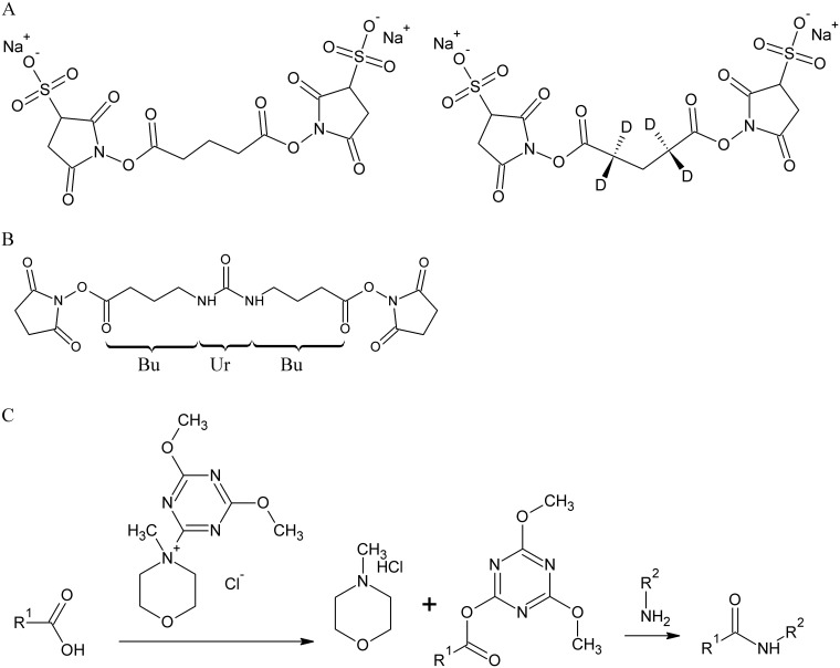
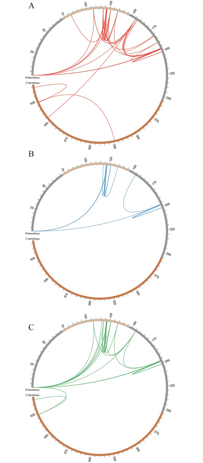
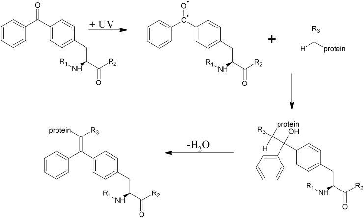
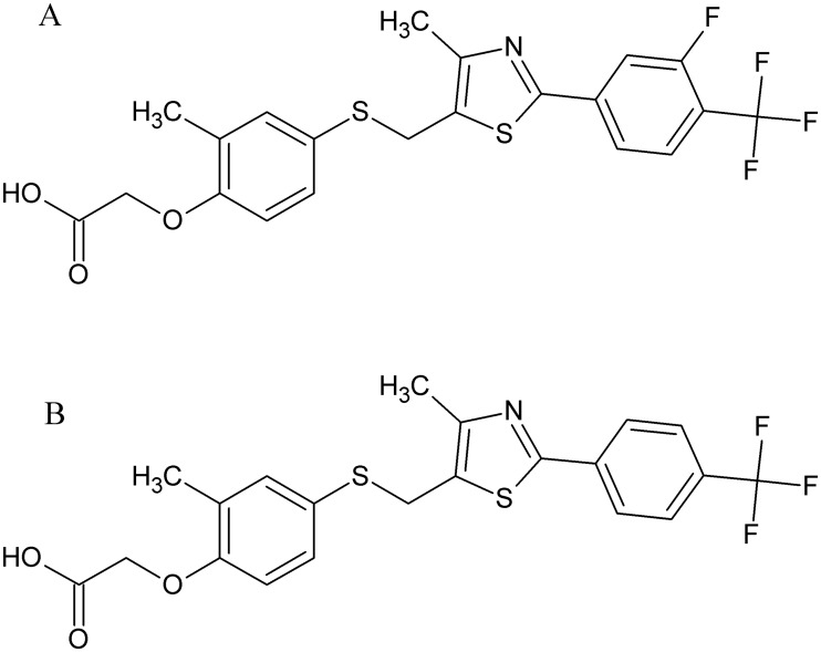
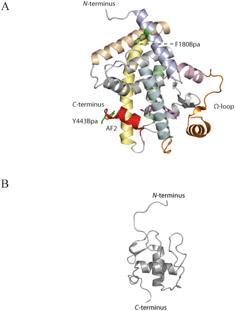
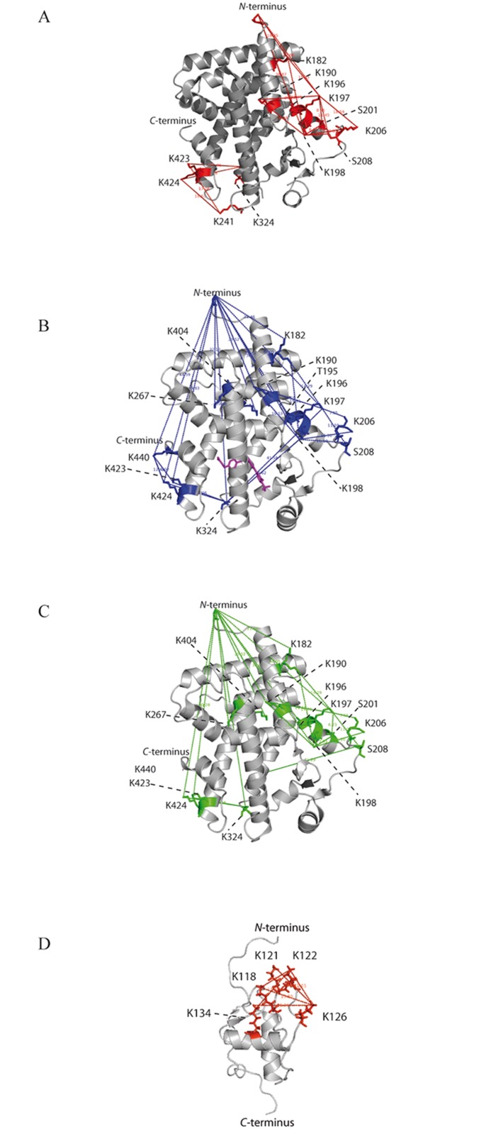
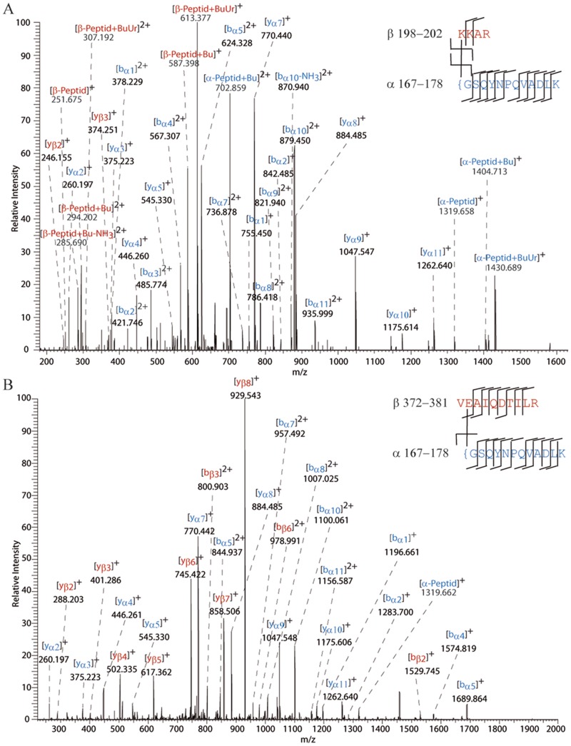
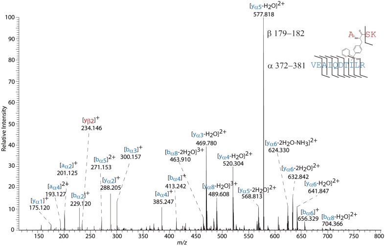
Peroxisome proliferator-activated receptors (PPARs) have been intensively studied as drug targets to treat type 2 diabetes, lipid disorders, and metabolic syndrome. This study is part of our ongoing efforts to map conformational changes in PPARs in solution by a combination of chemical cross-linking and mass spectrometry (MS). To our best knowledge, we performed the first studies addressing solution structures of full-length PPAR-β/δ. We monitored the conformations of the ligand-binding domain (LBD) as well as full-length PPAR-β/δ upon binding of two agonists. (Photo-) cross-linking relied on (i) a variety of externally introduced amine- and carboxyl-reactive linkers and (ii) the incorporation of the photo-reactive amino acid p-benzoylphenylalanine (Bpa) into PPAR-β/δ by genetic engineering. The distances derived from cross-linking experiments allowed us to monitor conformational changes in PPAR-β/δ upon ligand binding. The cross-linking/MS approach proved highly advantageous to study nuclear receptors, such as PPARs, and revealed the interplay between DBD (DNA-binding domain) and LDB in PPAR-β/δ. Our results indicate the stabilization of a specific conformation through ligand binding in PPAR-β/δ LBD as well as full-length PPAR-β/δ. Moreover, our results suggest a close distance between the N- and C-terminal regions of full-length PPAR-β/δ in the presence of GW1516. Chemical cross-linking/MS allowed us gaining detailed insights into conformational changes that are induced in PPARs when activating ligands are present. Thus, cross-linking/MS should be added to the arsenal of structural methods available for studying nuclear receptors.
过氧化物酶体增殖物激活受体(PPARs)作为治疗2型糖尿病、脂质紊乱和代谢综合征的药物靶点,已得到深入研究。本研究是我们通过化学交联和质谱(MS)相结合的方法绘制溶液中PPARs构象变化的持续努力的一部分。据我们所知,我们首次开展了关于全长PPAR-β/δ溶液结构的研究。我们监测了两种激动剂结合后配体结合域(LBD)以及全长PPAR-β/δ的构象。(光)交联依赖于(i)多种外部引入的胺反应性和羧基反应性连接子,以及(ii)通过基因工程将光反应性氨基酸对苯甲酰基苯丙氨酸(Bpa)掺入PPAR-β/δ中。交联实验得出的距离使我们能够监测配体结合后PPAR-β/δ的构象变化。交联/MS方法被证明在研究核受体(如PPARs)方面具有高度优势,并揭示了PPAR-β/δ中DNA结合域(DBD)和配体结合域(LDB)之间的相互作用。我们的结果表明,通过配体结合,PPAR-β/δ LBD以及全长PPAR-β/δ中的特定构象得以稳定。此外,我们的结果表明,在存在GW1516的情况下,全长PPAR-β/δ的N端和C端区域之间距离很近。化学交联/MS使我们能够深入了解激活配体存在时PPARs中诱导的构象变化。因此,交联/MS应被添加到可用于研究核受体的结构方法库中。