Lim Manyoel, Kim June Sic, Kim Dajung J, Chung Chun Kee
Neuroscience Research Institute, Seoul National University College of Medicine Seoul, South Korea.
Department of Brain and Cognitive Sciences, Seoul National University College of Natural Sciences Seoul, South Korea.
Front Hum Neurosci. 2016 Mar 14;10:111. doi: 10.3389/fnhum.2016.00111. eCollection 2016.
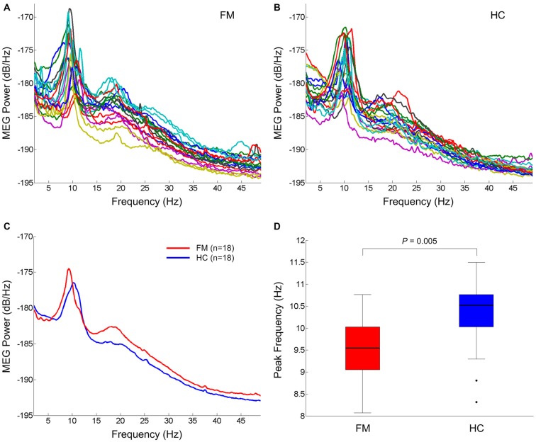
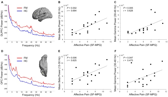
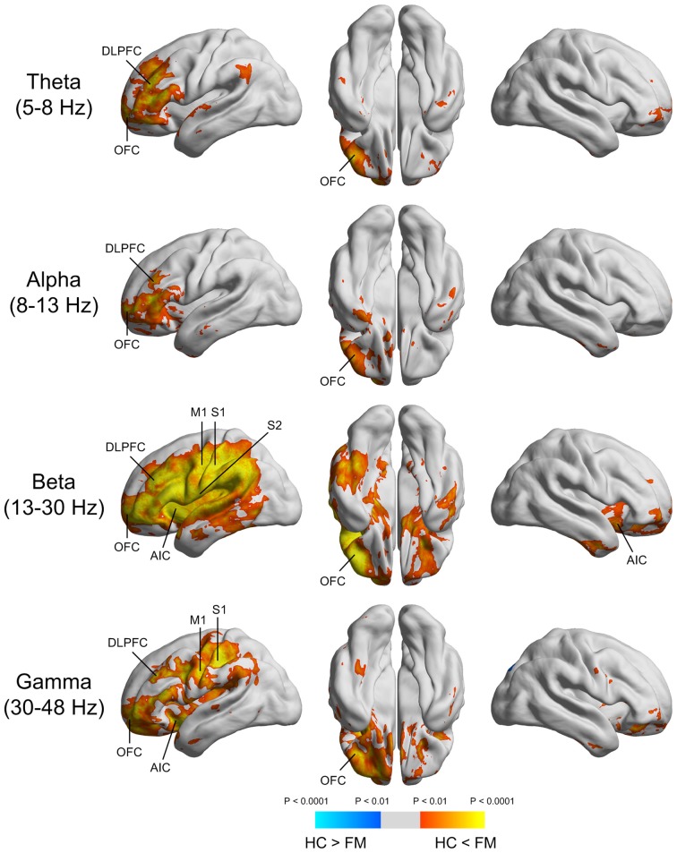
Recent human neuroimaging studies have suggested that fibromyalgia (FM), a chronic widespread pain disorder, exhibits altered thalamic structure and function. Since the thalamus has extensive reciprocal connection with the cortex, structural and functional thalamic alterations in FM might be linked to aberrant thalamocortical oscillation. This study investigated the presence of abnormal brain rhythmicity in low- and high-frequency bands during resting state in patients with FM and their relationship to clinical pain symptom. Spontaneous magnetoencephalography (MEG) activity was recorded in 18 females with FM and 18 age- and sex-matched healthy control (HC) subjects. The most remarkable finding was that FM patients had general increases in theta, beta and gamma power along with a slowing of the dominant alpha peak. Increased spectral powers in the theta-band were primarily localized to the left dorsolateral prefrontal (DLPFC) and orbitofrontal cortex (OFC). Beta and gamma over-activation were localized to insular, primary motor and primary and secondary somatosensory (S2) cortices, as well as the DLPFC and OFC. Furthermore, enhanced high-frequency oscillatory activities in the DLPFC and OFC were associated with higher affective pain scores in patients with FM. Our results demonstrate that FM patients feature enhanced low- and high-frequency oscillatory activity in the brain areas related to cognitive and emotional modulation of pain. Increased low- and high-frequency activity of the prefrontal cortex may contribute to persistent perception of pain in FM. Therapeutic intervention based on manipulating neural oscillation to restore normal thalamocortical rhythmicity may be beneficial to pain relief in FM.
近期的人体神经影像学研究表明,纤维肌痛(FM)作为一种慢性广泛性疼痛疾病,其丘脑结构和功能出现了改变。由于丘脑与皮层有着广泛的相互连接,FM患者丘脑的结构和功能改变可能与异常的丘脑皮质振荡有关。本研究调查了FM患者静息状态下低频和高频波段脑节律异常的情况及其与临床疼痛症状的关系。对18名患有FM的女性和18名年龄及性别匹配的健康对照(HC)受试者进行了自发性脑磁图(MEG)活动记录。最显著的发现是,FM患者的θ波、β波和γ波功率普遍增加,同时主导α波峰变慢。θ波段功率增加主要局限于左侧背外侧前额叶(DLPFC)和眶额皮质(OFC)。β波和γ波过度激活局限于岛叶、初级运动皮层以及初级和次级躯体感觉(S2)皮层,以及DLPFC和OFC。此外,DLPFC和OFC中高频振荡活动增强与FM患者更高的情感性疼痛评分相关。我们的结果表明,FM患者在与疼痛的认知和情绪调节相关的脑区呈现出增强的低频和高频振荡活动。前额叶皮层低频和高频活动增加可能导致FM患者持续的疼痛感知。基于操纵神经振荡以恢复正常丘脑皮质节律性的治疗干预可能有助于缓解FM患者的疼痛。