• 文献检索
  • 文档翻译
  • 深度研究
  • 学术资讯
  • Suppr Zotero 插件Zotero 插件
  • 邀请有礼
  • 套餐&价格
  • 历史记录
应用&插件
Suppr Zotero 插件Zotero 插件浏览器插件Mac 客户端Windows 客户端微信小程序
定价
高级版会员购买积分包购买API积分包
服务
文献检索文档翻译深度研究API 文档MCP 服务
关于我们
关于 Suppr公司介绍联系我们用户协议隐私条款
关注我们

Suppr 超能文献

核心技术专利:CN118964589B侵权必究
粤ICP备2023148730 号-1Suppr @ 2026

文献检索

告别复杂PubMed语法,用中文像聊天一样搜索,搜遍4000万医学文献。AI智能推荐,让科研检索更轻松。

立即免费搜索

文件翻译

保留排版,准确专业,支持PDF/Word/PPT等文件格式,支持 12+语言互译。

免费翻译文档

深度研究

AI帮你快速写综述,25分钟生成高质量综述,智能提取关键信息,辅助科研写作。

立即免费体验

肾动脉狭窄的超声诊断:狭窄标准、超声造影及支架内再狭窄

Ultrasound diagnostics of renal artery stenosis: Stenosis criteria, CEUS and recurrent in-stent stenosis.

作者信息

Schäberle W, Leyerer L, Schierling W, Pfister K

机构信息

Department of Visceral, Vascular, Thorax and Pediatric Surgery, "Klinik am Eichert", Eichertstr. 3, 73035 Göppingen, Germany.

Vascular and Endovascular Surgery, Regensburg University Hospital, Regensburg, Germany.

出版信息

Gefasschirurgie. 2016;21:4-13. doi: 10.1007/s00772-015-0060-3. Epub 2015 Aug 28.

DOI:10.1007/s00772-015-0060-3
PMID:27034579
原文链接:https://pmc.ncbi.nlm.nih.gov/articles/PMC4767855/
Abstract

BACKGROUND AND PURPOSE

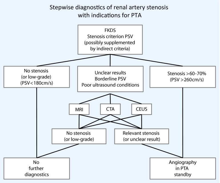
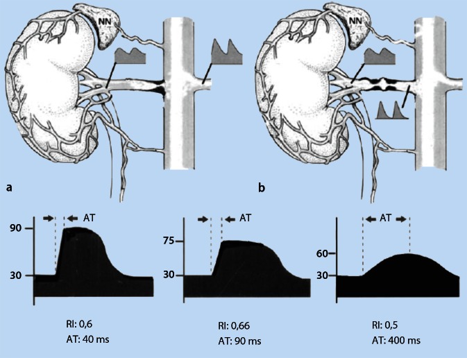
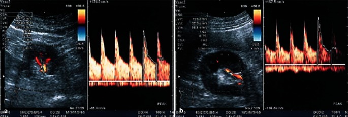
As a non-invasive, side effect-free and cost-effective method, ultrasonography represents the method of choice for the diagnosis of renal artery stenosis. Four different criteria in total, including two direct criteria in peak systolic velocity (PSV) and renal aortic ratio (RAR) and two indirect criteria in resistance index (RI) and acceleration time (AT) for the measurement of relevant renal artery stenosis are described, each demonstrating highly variable accuracy in studies. Furthermore, there is controversy over the degree beyond which stenosis becomes therapeutically relevant and which ultrasound PSV is diagnostically relevant in terms of stenosis grading.

MATERIAL AND METHODS

This article gives a critical review based on a selective literature search on measurement methodology and the validity of ultrasound in renal artery stenosis. A critical evaluation of methods and a presentation of measurement principles to establish the most precise measurement method possible compared with the gold standard angiography, as well as an evaluation of the importance of computed tomography angiography (CTA) and magnetic resonance angiography (MRA).

RESULTS AND CONCLUSIONS

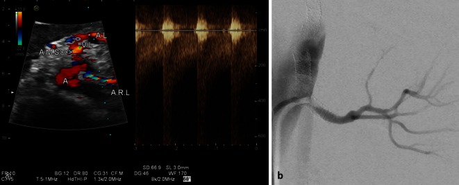
The PSV provides high sensitivity and specificity as a direct measurement method in stenosis detection and grading. Most studies found sensitivities and specificities of 85-90 % for > 50 % stenosis at a PSV > 180-200 cm/s in ROC curve analysis. Other methods, such as the ratio of the PSV in the aorta to the PSV in the renal artery (RAR) or indirect criteria, such as side to side differences in RI (dRI) or AT can be additionally used to improve accuracy. Contrast-enhanced ultrasound improves accuracy by means of echo contrast enhancement. Although in the past only high-grade stenosis was considered relevant for treatment, a drop in pressure of > 20 mmHg in > 50 % stenosis (PSV 180 cm/s) is classified as relevant for increased renin secretion. Stenosis in fibromuscular dysplasia can be reliably graded according to the continuity equation. Although the available studies on the grading of in-stent restenosis are the subject of controversy, there is a tendency to assume higher cut-off values for PSV and RAR. Whilst MRA and CTA demonstrate an accuracy of > 90 %, this is at the cost of possible side effects for patients, particularly in the case of pre-existing renal parenchymal damage.

ADDITIONAL ONLINE MATERIAL

This article includes two additional video sequences on visualizing renal artery stenosis. This supplemental material can be found under: dx.doi.org/10.1007/s00772-015-0060-3.

摘要

背景与目的

超声检查作为一种无创、无副作用且经济高效的方法,是诊断肾动脉狭窄的首选方法。总共描述了四种不同的标准,包括收缩期峰值流速(PSV)和肾主动脉比率(RAR)这两个直接标准,以及阻力指数(RI)和加速时间(AT)这两个间接标准,用于测量相关的肾动脉狭窄,每项标准在研究中的准确性都有很大差异。此外,对于狭窄在何种程度上具有治疗相关性以及在狭窄分级方面何种超声PSV具有诊断相关性存在争议。

材料与方法

本文基于对肾动脉狭窄测量方法及超声有效性的选择性文献检索进行批判性综述。对方法进行批判性评估,并介绍测量原理,以建立与金标准血管造影相比尽可能精确的测量方法,同时评估计算机断层血管造影(CTA)和磁共振血管造影(MRA)的重要性。

结果与结论

PSV作为狭窄检测和分级的直接测量方法具有高灵敏度和特异性。大多数研究在ROC曲线分析中发现,对于PSV>180 - 200 cm/s时>50%的狭窄,灵敏度和特异性为85 - 90%。其他方法,如主动脉PSV与肾动脉PSV的比率(RAR)或间接标准,如RI的左右差异(dRI)或AT,可额外用于提高准确性。超声造影通过增强回声对比提高准确性。尽管过去仅认为高度狭窄与治疗相关,但对于>50%狭窄(PSV 180 cm/s)时压力下降>20 mmHg,归类为与肾素分泌增加相关。纤维肌发育不良中的狭窄可根据连续性方程可靠分级。尽管关于支架内再狭窄分级的现有研究存在争议,但倾向于采用更高的PSV和RAR截断值。虽然MRA和CTA的准确性>90%,但这是以患者可能出现副作用为代价的,尤其是在已有肾实质损害的情况下。

附加在线材料

本文包括两段关于可视化肾动脉狭窄的附加视频序列。该补充材料可在以下网址找到:dx.doi.org/10.1007/s00772-015-0060-3。

https://cdn.ncbi.nlm.nih.gov/pmc/blobs/d8e6/4767855/dc4b10439760/772_2015_60_Fig8_HTML.jpg
https://cdn.ncbi.nlm.nih.gov/pmc/blobs/d8e6/4767855/d3ea62845418/772_2015_60_Fig1_HTML.jpg
https://cdn.ncbi.nlm.nih.gov/pmc/blobs/d8e6/4767855/3645a84061fd/772_2015_60_Fig2_HTML.jpg
https://cdn.ncbi.nlm.nih.gov/pmc/blobs/d8e6/4767855/b5cff2f7f854/772_2015_60_Fig3_HTML.jpg
https://cdn.ncbi.nlm.nih.gov/pmc/blobs/d8e6/4767855/f262cc700851/772_2015_60_Fig4_HTML.jpg
https://cdn.ncbi.nlm.nih.gov/pmc/blobs/d8e6/4767855/8a60646e05cf/772_2015_60_Fig5_HTML.jpg
https://cdn.ncbi.nlm.nih.gov/pmc/blobs/d8e6/4767855/6227af37cf49/772_2015_60_Fig6_HTML.jpg
https://cdn.ncbi.nlm.nih.gov/pmc/blobs/d8e6/4767855/5ee23dd139c7/772_2015_60_Fig7_HTML.jpg
https://cdn.ncbi.nlm.nih.gov/pmc/blobs/d8e6/4767855/dc4b10439760/772_2015_60_Fig8_HTML.jpg

相似文献

1
Ultrasound diagnostics of renal artery stenosis: Stenosis criteria, CEUS and recurrent in-stent stenosis.肾动脉狭窄的超声诊断:狭窄标准、超声造影及支架内再狭窄
Gefasschirurgie. 2016;21:4-13. doi: 10.1007/s00772-015-0060-3. Epub 2015 Aug 28.
2
Critical analysis of renal duplex ultrasound parameters in detecting significant renal artery stenosis.对肾双功超声参数在检测显著肾动脉狭窄中的作用进行分析。
J Vasc Surg. 2012 Oct;56(4):1052-9, 1060.e1; discussion 1059-60. doi: 10.1016/j.jvs.2012.03.036. Epub 2012 May 15.
3
Duplex ultrasound velocity criteria for the stented carotid artery.支架置入后颈动脉的双功超声速度标准。
J Vasc Surg. 2008 Jan;47(1):63-73. doi: 10.1016/j.jvs.2007.09.038.
4
Renal duplex ultrasound findings in fenestrated endovascular aortic repair for juxtarenal aortic aneurysms.开窗式血管腔内主动脉修复术治疗近肾腹主动脉瘤的肾双功超声检查结果
J Vasc Surg. 2016 Apr;63(4):915-21. doi: 10.1016/j.jvs.2015.10.090. Epub 2015 Dec 31.
5
Renal artery duplex ultrasound criteria for the detection of significant in-stent restenosis.检测显著支架内再狭窄的肾动脉双功超声标准。
Catheter Cardiovasc Interv. 2014 Mar 1;83(4):612-8. doi: 10.1002/ccd.25270. Epub 2013 Nov 14.
6
Best duplex-sonographic criteria for the assessment of renal artery stenosis--correlation with intra- arterial pressure gradient.评估肾动脉狭窄的最佳双功超声标准——与动脉内压力梯度的相关性
Ultraschall Med. 2007 Feb;28(1):45-51. doi: 10.1055/s-2007-962881.
7
Optimal carotid duplex velocity criteria for defining the severity of carotid in-stent restenosis.用于定义颈动脉支架内再狭窄严重程度的最佳颈动脉双功超声速度标准。
J Vasc Surg. 2008 Sep;48(3):589-94. doi: 10.1016/j.jvs.2008.04.004. Epub 2008 Jun 30.
8
Predictive multivariate regression to increase the specificity of carotid duplex ultrasound for high-grade stenosis in asymptomatic patients.预测性多变量回归以提高颈动脉双功超声对无症状患者重度狭窄的特异性。
Ann Vasc Surg. 2014 Aug;28(6):1548-55. doi: 10.1016/j.avsg.2014.02.010. Epub 2014 Feb 12.
9
Determining in-stent stenosis of carotid arteries by duplex ultrasound criteria.通过双功超声标准确定颈动脉支架内狭窄情况。
J Endovasc Ther. 2005 Jun;12(3):346-53. doi: 10.1583/04-1527.1.
10
The performance of duplex ultrasonography for the assessment of renal artery stenosis in Takayasu's arteritis patients.双功能超声在评估多发性大动脉炎患者肾动脉狭窄中的应用。
Arthritis Res Ther. 2023 Aug 3;25(1):139. doi: 10.1186/s13075-023-03121-8.

引用本文的文献

1
Assessment of renal artery stenosis using renal fractional flow reserve and correlation with angiography and color Doppler ultrasonography: data from FAIR-pilot trial.使用肾血流储备分数评估肾动脉狭窄及其与血管造影和彩色多普勒超声检查的相关性:来自FAIR试点试验的数据。
Hypertens Res. 2025 Feb;48(2):702-709. doi: 10.1038/s41440-024-01948-5. Epub 2024 Oct 21.
2
Management of Renovascular Hypertension and Renal Denervation in Patients with Hypertension: An Italian Nationwide Survey.高血压患者的肾血管性高血压和肾去神经治疗管理:意大利全国性调查。
High Blood Press Cardiovasc Prev. 2024 Sep;31(5):501-512. doi: 10.1007/s40292-024-00668-8. Epub 2024 Sep 18.
3

本文引用的文献

1
Critical analysis of renal duplex ultrasound parameters in detecting significant renal artery stenosis.对肾双功超声参数在检测显著肾动脉狭窄中的作用进行分析。
J Vasc Surg. 2012 Oct;56(4):1052-9, 1060.e1; discussion 1059-60. doi: 10.1016/j.jvs.2012.03.036. Epub 2012 May 15.
2
Comparison of duplex ultrasonography and magnetic resonance imaging in the detection of significant renal artery stenosis.双功超声检查与磁共振成像在检测显著肾动脉狭窄中的比较。
Acta Medica (Hradec Kralove). 2011;54(1):9-12. doi: 10.14712/18059694.2016.10.
3
The clinical role of contrast-enhanced ultrasound in the evaluation of renal artery stenosis and diagnostic superiority as compared to traditional echo-color-Doppler flow imaging.
Impact of Breathing Control Training Program on sonographic quantification of abdominal vasculature.
呼吸控制训练方案对腹部血管超声量化的影响。
Saudi Med J. 2024 May;45(5):525-530. doi: 10.15537/smj.2024.45.5.20240083.
4
The performance of duplex ultrasonography for the assessment of renal artery stenosis in Takayasu's arteritis patients.双功能超声在评估多发性大动脉炎患者肾动脉狭窄中的应用。
Arthritis Res Ther. 2023 Aug 3;25(1):139. doi: 10.1186/s13075-023-03121-8.
5
Case Report: Endovascular Treatment of Chronic Atherosclerotic Renal Artery Total Occlusions with Failed Medical Therapy.病例报告:内科治疗失败的慢性动脉粥样硬化性肾动脉完全闭塞的血管内治疗
Front Surg. 2022 Oct 18;9:843568. doi: 10.3389/fsurg.2022.843568. eCollection 2022.
6
Vascular imaging for anaesthetists and intensivists, part 1: Basic principles.麻醉医师和重症监护医师的血管成像,第1部分:基本原则。
BJA Educ. 2022 Sep;22(9):334-342. doi: 10.1016/j.bjae.2022.04.003. Epub 2022 Jul 16.
7
Gray-Scale, Color Doppler, Spectral Doppler, and Contrast-Enhanced Renal Artery Ultrasound: Imaging Techniques and Features.灰阶、彩色多普勒、频谱多普勒及超声造影增强肾动脉超声:成像技术与特征
J Clin Med. 2022 Jul 7;11(14):3961. doi: 10.3390/jcm11143961.
8
Value of contrast-enhanced ultrasound for diagnosis and follow-up of renal artery stenosis in patients with chronic kidney disease.超声造影在慢性肾脏病患者肾动脉狭窄诊断及随访中的价值
Abdom Radiol (NY). 2022 May;47(5):1853-1861. doi: 10.1007/s00261-022-03457-w. Epub 2022 Mar 5.
9
The Value of Contrast-Enhanced Ultrasound versus Doppler Ultrasound in Grading Renal Artery Stenosis.对比增强超声与多普勒超声在肾动脉狭窄分级中的价值。
Biomed Res Int. 2020 Sep 8;2020:7145728. doi: 10.1155/2020/7145728. eCollection 2020.
10
Renal Artery Stenosis in the Patient with Hypertension: Prevalence, Impact and Management.高血压患者的肾动脉狭窄:患病率、影响及管理
Integr Blood Press Control. 2020 Jun 2;13:71-82. doi: 10.2147/IBPC.S248579. eCollection 2020.
超声造影在评估肾动脉狭窄中的临床作用以及与传统超声彩色多普勒血流成像相比的诊断优势。
Int Angiol. 2011 Apr;30(2):135-9.
4
Accuracy of duplex sonography scans after renal artery stenting.肾动脉支架置入术后的双功能超声扫描的准确性。
J Vasc Surg. 2010 Oct;52(4):953-7; discussion 958. doi: 10.1016/j.jvs.2010.04.055.
5
[Ultrasound diagnostics of renal blood vessels and transplant kidney].
Radiologe. 2009 Nov;49(11):1040-7. doi: 10.1007/s00117-009-1873-4.
6
64-Detector CT angiography in renal artery stent evaluation: prospective comparison with selective catheter angiography.
Radiology. 2009 Jul;252(1):299-305. doi: 10.1148/radiol.2521081362.
7
Ultrasound velocity criteria for renal in-stent restenosis.肾支架内再狭窄的超声速度标准。
J Vasc Surg. 2009 Jul;50(1):119-23. doi: 10.1016/j.jvs.2008.12.066. Epub 2009 Feb 23.
8
Renal artery stenting with noninvasive duplex ultrasound follow-up: 3-year results from the RENAISSANCE renal stent trial.肾动脉支架置入术联合无创双功超声随访:RENAISSANCE肾支架试验的3年结果
Catheter Cardiovasc Interv. 2008 Nov 15;72(6):853-62. doi: 10.1002/ccd.21749.
9
Imaging modalities for renal artery stenosis in suspected renovascular hypertension: prospective intraindividual comparison of color Doppler US, CT angiography, GD-enhanced MR angiography, and digital substraction angiography.疑似肾血管性高血压患者肾动脉狭窄的成像方式:彩色多普勒超声、CT血管造影、钆增强磁共振血管造影和数字减影血管造影的前瞻性个体内比较
Ren Fail. 2007;29(3):295-302. doi: 10.1080/08860220601166305.
10
Best duplex-sonographic criteria for the assessment of renal artery stenosis--correlation with intra- arterial pressure gradient.评估肾动脉狭窄的最佳双功超声标准——与动脉内压力梯度的相关性
Ultraschall Med. 2007 Feb;28(1):45-51. doi: 10.1055/s-2007-962881.