Pal Madan, Singh Prem, Tayal Rishi, Dehmiwal Dinesh, Behl S M, Kumar Sarvan, Chandolia R K
Department of Veterinary Surgery and Radiology, Lala Lajpat Rai University of Veterinary & Animal Sciences, Hisar, Haryana, India.
Department of Veterinary Pathology, Lala Lajpat Rai University of Veterinary & Animal Sciences, Hisar, Haryana, India.
Vet World. 2015 Jun;8(6):707-12. doi: 10.14202/vetworld.2015.707-712. Epub 2015 Jun 6.
The objective of the study was to obtain and compare the two-dimensional (2D) and three-dimensional (3D) ultrasonographic images of pathological conditions of the stomach in dogs in clinical cases.
In our study, 12 clinical conditions of the stomach were recorded using ultrasonography. The ultrasound machine used for this study was 3D ultrasound machine (Nemio-XG: Toshiba, Japan) having four-dimensional volumetric transducer.
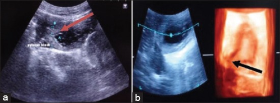
Present study was done to compare 2D and 3D ultrasonographic images in different gastric affections in dogs. In case of uremic gastropathy due to inflammatory response, the wall of the stomach was 0.6 cm thick and hyperechoic and gastric folds were also hyperechoic indicative of gastritis. In second, third, and fourth case of gastritis the wall of the stomach was 0.7, 0.6, and 0.55 cm, respectively thick and hyperechoic. In fifth and sixth case of gastritis, inflammatory response due to ingestion of polythene and sand led to gastritis and ultrasonographically, the wall of the stomach was 0.6 cm and 0.7 cm thick, respectively, and hyperechoic. In case of gastric ulcer, ultrasonographically, there was a disruption of gastric mucosal layer. In cases of gastric dilatation, anechoic content indicating fluid was seen in stomach area and due to dilatation boundary of the stomach was not clear and the increase in the lumen of the stomach was observed. In case of foreign body, ultrasonographically the wall of the stomach was 0.55 cm thick and hyperechoic. In the middle of the stomach, multiple hyperechoic shadows of the foreign bodies i.e. leather and bunch of straw of grass were observed. In case of pyloric stenosis ultrasonographically, anechoic lumen of the pylorus surrounded by 0.5 cm hypoechoic thickened muscle. In some cases, 3D ultrasonography was not diagnostic i.e. gastric foreign bodies and gastric dilatation. These conditions were better visualized on the 2D sonogram.
The appearance of clinical conditions of the stomach such as gastritis and pyloric stenosis were more distinct on 3D ultrasonogram than 2D ultrasonogram. The 3D ultrasonography was not diagnostic in cases of gastric foreign bodies and gastric dilatation.
本研究的目的是获取并比较临床病例中犬胃病理状况的二维(2D)和三维(3D)超声图像。
在我们的研究中,使用超声检查记录了12例胃的临床状况。本研究使用的超声仪是具有四维容积探头的3D超声仪(Nemio-XG:日本东芝)。
本研究旨在比较犬不同胃部疾病中的2D和3D超声图像。在因炎症反应导致的尿毒症性胃病中,胃壁厚度为0.6厘米,呈高回声,胃皱襞也呈高回声,提示胃炎。在第二、第三和第四例胃炎中,胃壁厚度分别为0.7、0.6和0.55厘米,呈高回声。在第五和第六例胃炎中,因摄入聚乙烯和沙子导致的炎症反应引发了胃炎,超声检查显示胃壁厚度分别为0.6厘米和0.7厘米,呈高回声。在胃溃疡病例中,超声检查显示胃黏膜层中断。在胃扩张病例中,在胃区域可见无回声内容物,提示有液体,由于扩张,胃的边界不清晰,且观察到胃腔增大。在异物病例中,超声检查显示胃壁厚度为0.55厘米,呈高回声。在胃中部,观察到多个异物的高回声阴影,即皮革和一束稻草。在幽门狭窄病例中,超声检查显示幽门的无回声管腔被0.5厘米低回声增厚肌肉包围。在某些情况下,3D超声检查无法做出诊断,即胃异物和胃扩张。这些情况在2D超声图上显示得更好。
胃炎和幽门狭窄等胃临床状况在3D超声图上的表现比2D超声图更清晰。3D超声检查在胃异物和胃扩张病例中无法做出诊断。