de Fatima Vasco Aragao Maria, van der Linden Vanessa, Brainer-Lima Alessandra Mertens, Coeli Regina Ramos, Rocha Maria Angela, Sobral da Silva Paula, Durce Costa Gomes de Carvalho Maria, van der Linden Ana, Cesario de Holanda Arthur, Valenca Marcelo Moraes
Centro Diagnostico Multimagem, Rua Frei Matias Tevis, 194, Ilha do Leite Recife Pernambuco 52010-450, Brazil; Medical School, Mauricio de Nassau University, Recife, Pernambuco, Brazil
Association for Assistance of Disabled Children, Recife, Brazil; Barão de Lucena Hospital, Recife, Brazil.
BMJ. 2016 Apr 13;353:i1901. doi: 10.1136/bmj.i1901.
To report radiological findings observed in computed tomography (CT) and magnetic resonance imaging (MRI) scans of the first cases of congenital infection and microcephaly presumably associated with the Zika virus in the current Brazilian epidemic.
Retrospective study with a case series.
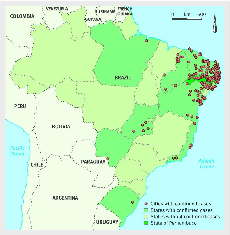
Association for Assistance of Disabled Children (AACD), Pernambuco state, Brazil.
23 children with a diagnosis of congenital infection presumably associated with the Zika virus during the Brazilian microcephaly epidemic.
Types of abnormalities and the radiological pattern of lesions identified on CT and MRI brain scans.
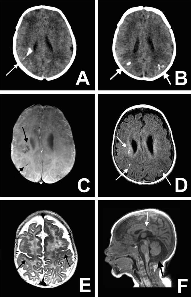
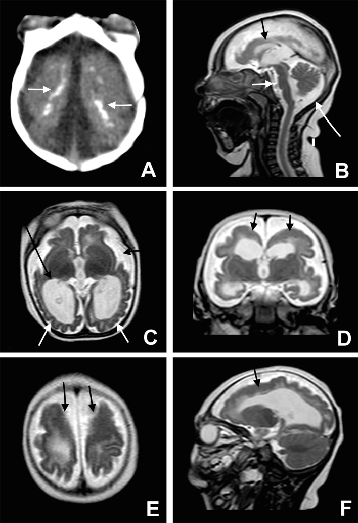
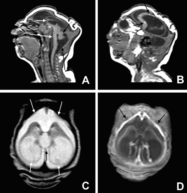
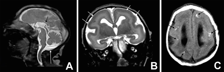
Six of the 23 children tested positive for IgM antibodies to Zika virus in cerebrospinal fluid. The other 17 children met the protocol criteria for congenital infection presumably associated with the Zika virus, even without being tested for IgM antibodies to the virus--the test was not yet available on a routine basis. Of the 23 children, 15 underwent CT, seven underwent both CT and MRI, and one underwent MRI. Of the 22 children who underwent CT, all had calcifications in the junction between cortical and subcortical white matter, 21 (95%) had malformations of cortical development, 20 (91%) had a decreased brain volume, 19 (86%) had ventriculomegaly, and 11 (50%) had hypoplasia of the cerebellum or brainstem. Of the eight children who underwent MRI, all had calcifications in the junction between cortical and subcortical white matter, malformations of cortical development occurring predominantly in the frontal lobes, and ventriculomegaly. Seven of the eight (88%) children had enlarged cisterna magna, seven (88%) delayed myelination, and six each (75%) a moderate to severe decrease in brain volume, simplified gyral pattern, and abnormalities of the corpus callosum (38% hypogenesis and 38% hypoplasia). Malformations were symmetrical in 75% of the cases.
Severe cerebral damage was found on imaging in most of the children in this case series with congenital infection presumably associated with the Zika virus. The features most commonly found were brain calcifications in the junction between cortical and subcortical white matter associated with malformations of cortical development, often with a simplified gyral pattern and predominance of pachygyria or polymicrogyria in the frontal lobes. Additional findings were enlarged cisterna magna, abnormalities of corpus callosum (hypoplasia or hypogenesis), ventriculomegaly, delayed myelination, and hypoplasia of the cerebellum and the brainstem.
报告在计算机断层扫描(CT)和磁共振成像(MRI)扫描中观察到的,与当前巴西疫情中寨卡病毒相关的首批先天性感染和小头畸形病例的影像学表现。
病例系列回顾性研究。
巴西伯南布哥州残疾儿童援助协会(AACD)。
23名在巴西小头畸形疫情期间被诊断为可能与寨卡病毒相关的先天性感染儿童。
CT和MRI脑部扫描中识别出的异常类型及病变的影像学特征。
23名儿童中,6名脑脊液中寨卡病毒IgM抗体检测呈阳性。另外17名儿童符合可能与寨卡病毒相关的先天性感染的方案标准,即便未进行该病毒IgM抗体检测——该检测尚未常规开展。23名儿童中,15名接受了CT检查,7名同时接受了CT和MRI检查,1名接受了MRI检查。在接受CT检查的22名儿童中,所有人在皮质与皮质下白质交界处均有钙化,21名(95%)有皮质发育畸形,20名(91%)脑容量减小,19名(86%)有脑室扩大,11名(50%)有小脑或脑干发育不全。在接受MRI检查的8名儿童中,所有人在皮质与皮质下白质交界处均有钙化,皮质发育畸形主要发生在额叶,且有脑室扩大。8名儿童中有7名(88%)有小脑延髓池扩大,7名(88%)有髓鞘形成延迟现象,6名儿童(75%)各有脑容量中度至重度减小、脑回模式简化以及胼胝体异常(38%发育不全和38%发育不良)。75%的病例中畸形为对称性。
在该病例系列中,大多数可能与寨卡病毒相关的先天性感染儿童经影像学检查发现有严重脑损伤。最常见的特征是皮质与皮质下白质交界处的脑钙化,伴有皮质发育畸形,常伴有脑回模式简化,额叶多为巨脑回或多小脑回。其他表现包括小脑延髓池扩大、胼胝体异常(发育不全或发育不良)、脑室扩大、髓鞘形成延迟以及小脑和脑干发育不全。