Institute of Structural and Molecular Biology, Division of Biosciences, University College London, London, United Kingdom.
The Francis Crick Institute, Mill Hill Laboratory, London, United Kingdom.
PLoS One. 2016 Apr 21;11(4):e0154176. doi: 10.1371/journal.pone.0154176. eCollection 2016.
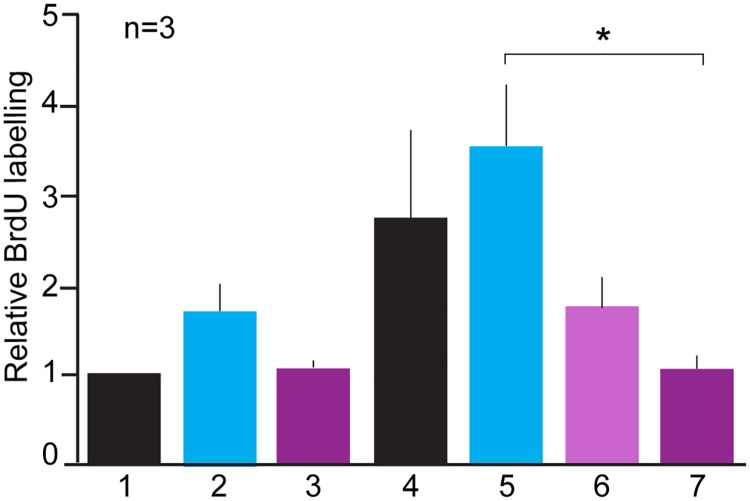
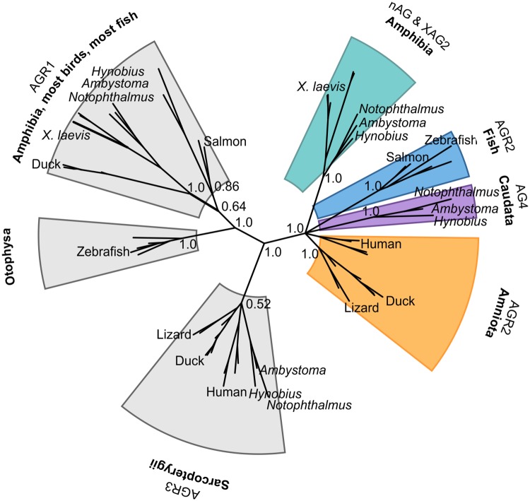
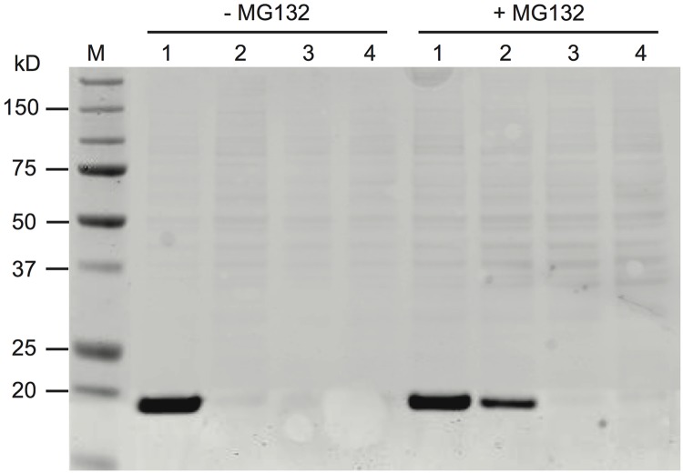
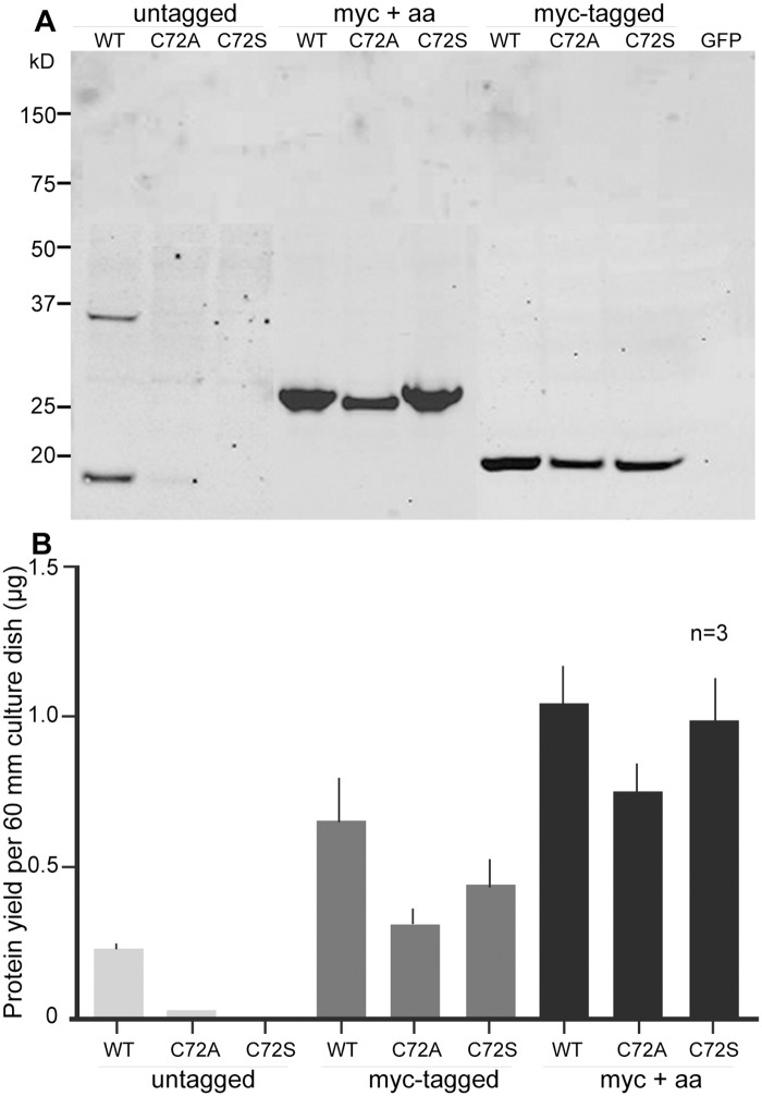
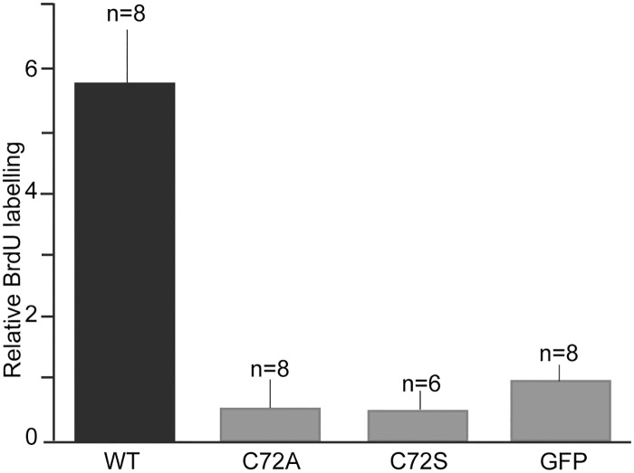
Anterior gradient (AG) proteins have a thioredoxin fold and are targeted to the secretory pathway where they may act in the ER, as well as after secretion into the extracellular space. A newt member of the family (nAG) was previously identified as interacting with the GPI-anchored salamander-specific three-finger protein called Prod1. Expression of nAG has been implicated in the nerve dependence of limb regeneration in salamanders, and nAG acted as a growth factor for cultured newt limb blastemal (progenitor) cells, but the mechanism of action was not understood. Here we show that addition of a peptide antibody to Prod1 specifically inhibit the proliferation of blastema cells, suggesting that Prod1 acts as a cell surface receptor for secreted nAG, leading to S phase entry. Mutation of the single cysteine residue in the canonical active site of nAG to alanine or serine leads to protein degradation, but addition of residues at the C terminus stabilises the secreted protein. The mutation of the cysteine residue led to no detectable activity on S phase entry in cultured newt limb blastemal cells. In addition, our phylogenetic analyses have identified a new Caudata AG protein called AG4. A comparison of the AG proteins in a cell culture assay indicates that nAG secretion is significantly higher than AGR2 or AG4, suggesting that this property may vary in different members of the family.
前梯度 (AG) 蛋白具有硫氧还蛋白折叠结构,靶向分泌途径,在那里它们可能在 ER 中发挥作用,以及在分泌到细胞外空间后发挥作用。先前已经鉴定出一种蝾螈家族的新成员(nAG)与 GPI 锚定的蝾螈特异性三指蛋白 Prod1 相互作用。nAG 的表达与蝾螈肢体再生的神经依赖性有关,nAG 作为培养的蝾螈肢体芽基(祖细胞)细胞的生长因子发挥作用,但作用机制尚不清楚。在这里,我们表明添加针对 Prod1 的肽抗体特异性抑制芽基细胞的增殖,表明 Prod1 作为分泌的 nAG 的细胞表面受体发挥作用,导致 S 期进入。将 nAG 典型活性位点中的单个半胱氨酸残基突变为丙氨酸或丝氨酸会导致蛋白降解,但 C 末端添加残基会稳定分泌蛋白。该半胱氨酸残基的突变导致在培养的蝾螈肢体芽基细胞中 S 期进入没有可检测到的活性。此外,我们的系统发育分析鉴定出一种新的有尾两栖类 AG 蛋白,称为 AG4。在细胞培养测定中比较 AG 蛋白表明,nAG 的分泌显著高于 AGR2 或 AG4,这表明该特性在家族的不同成员中可能有所不同。