Suppr超能文献

皮肤镜下扁平苔藓样角化病特征的时间顺序:17例总结

Chronology of lichen planus-like keratosis features by dermoscopy: a summary of 17 cases.

作者信息

Watanabe Soko, Sawada Mizuki, Dekio Itaru, Ishizaki Sumiko, Fujibayashi Mariko, Tanaka Masaru

机构信息

Department of Dermatology, Tokyo Women's Medical University Medical Center East, Tokyo, Japan.

Department of Pathology, Tokyo Women's Medical University Medical Center East, Tokyo, Japan.

出版信息

Dermatol Pract Concept. 2016 Apr 30;6(2):29-35. doi: 10.5826/dpc.0602a06. eCollection 2016 Apr.

Abstract

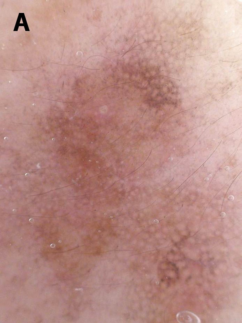
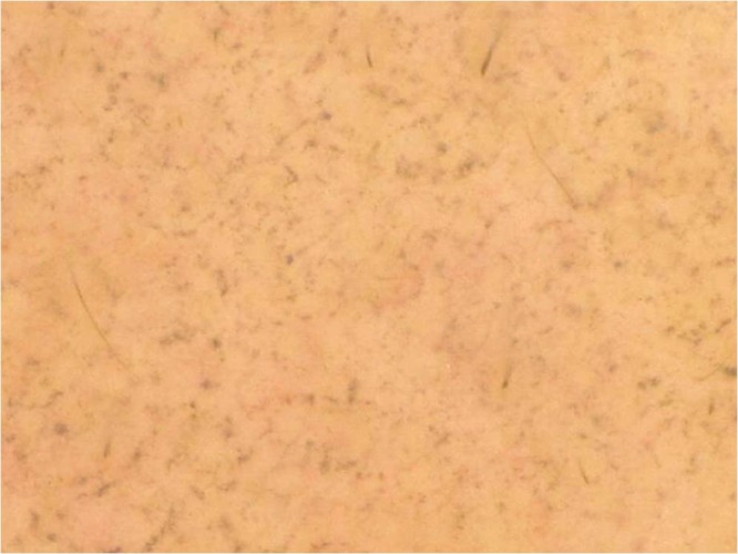
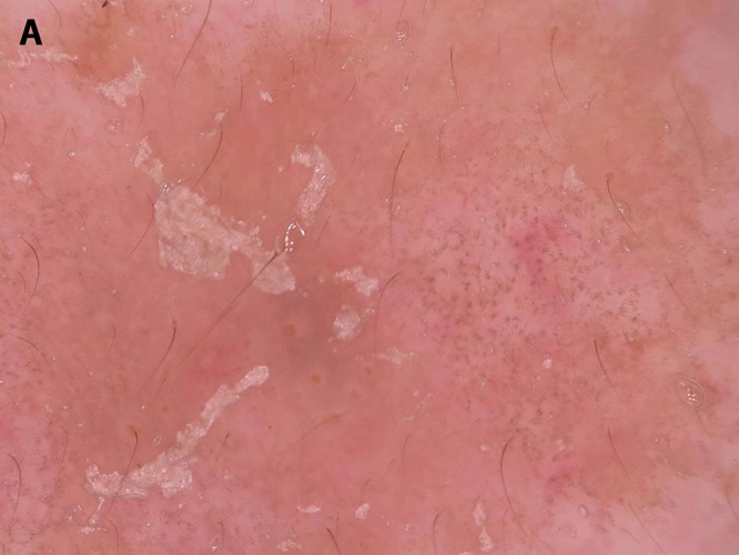
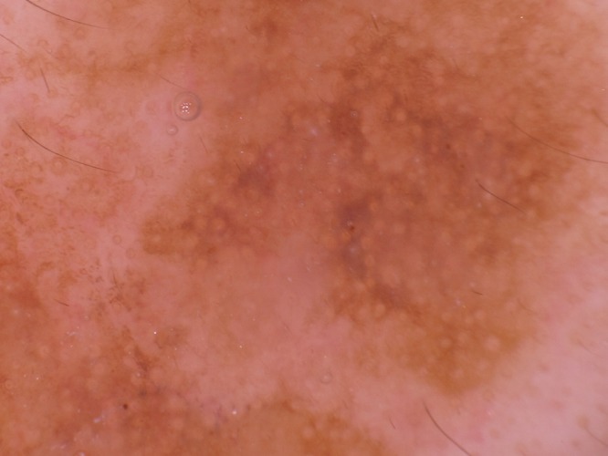
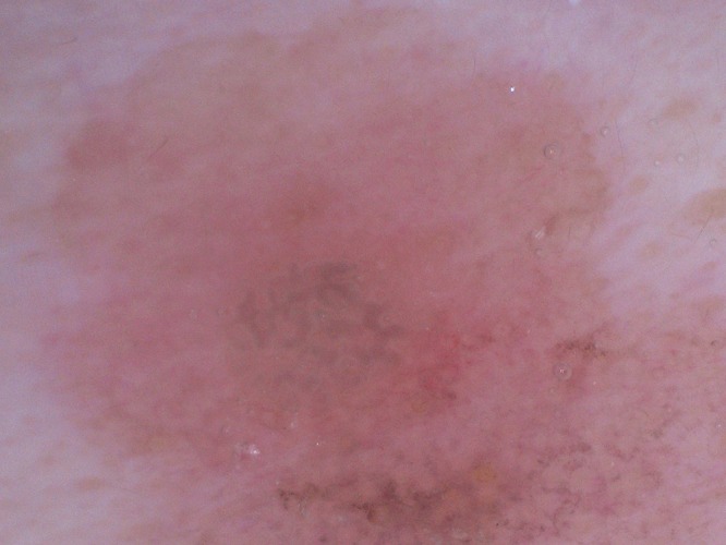
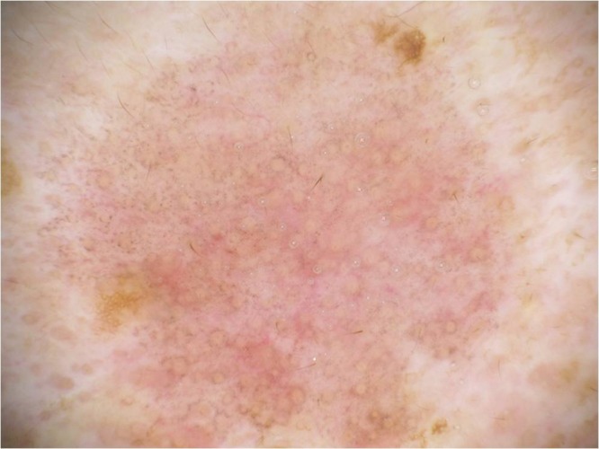
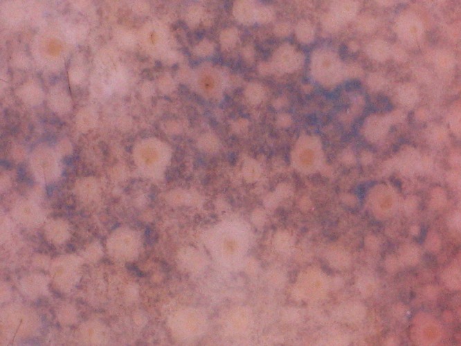
Dermoscopic findings for 17 cases of lichen planus-like keratosis (LPLK) were chronologically evaluated. Three males and 14 females were included in the study and the ages ranged from 43 to 85 years (median 65 years). Three cases were diagnosed based on stereotypical dermoscopic findings, while the other 14 cases were histopathologically diagnosed as LPLK. Dermoscopy photographs were divided into four groups depending on the number of days (D) from the initial visit: 1) D = 0 (initial visit or biopsy day); 2) D = 61 to 180; 3) D = 181 to 270; 4) D = 271 to 360. Dermoscopic findings, described as light brown pseudonetwork, pinkish area, gray pseudonetwork, annular granular structures, and blue-gray fine dots, were evaluated at every visit to the hospital. Initial dermoscopy features included light brown pseudonetworks due to residual solar lentigo and overlapping pinkish areas attributed to lichenoid inflammation. Annular granular structures and gray pseudonetwork appeared to be the main features of the regressing stage; these features seemed to progress to "blue-gray fine dots" in the late regressing stage. Blue-gray dots or globules reflecting melanophages, the hallmark dermoscopic features of LPLK, were believed to resolve in approximately one to two years. Based on the clinical and dermoscopic observations, we have specified five stages of evolution of LPLK, namely 1) pre-existing solar lentigo, 2) early inflammatory stage, 3) early regressing stage, 4) regressing stage, and 5) late regressing stage. The limitations of the study are that this is a small-sized, retrospective, observational study and that ethnicity of participants is limited to Japanese patients with skin phototype III.

摘要

对17例扁平苔藓样角化病(LPLK)的皮肤镜检查结果进行了时间顺序评估。该研究纳入了3名男性和14名女性,年龄范围为43至85岁(中位数65岁)。3例根据典型的皮肤镜检查结果确诊,另外14例经组织病理学诊断为LPLK。根据初次就诊后的天数(D),将皮肤镜照片分为四组:1)D = 0(初次就诊或活检日);2)D = 61至180;3)D = 181至270;4)D = 271至360。每次到医院就诊时,对描述为浅棕色假网络、粉红色区域、灰色假网络、环状颗粒结构和蓝灰色细点的皮肤镜检查结果进行评估。最初的皮肤镜特征包括残留的日光性雀斑样痣导致的浅棕色假网络以及苔藓样炎症引起的重叠粉红色区域。环状颗粒结构和灰色假网络似乎是消退期的主要特征;这些特征在消退后期似乎发展为“蓝灰色细点”。反映噬黑素细胞的蓝灰色点或小球是LPLK的标志性皮肤镜特征,据信在大约一到两年内会消退。基于临床和皮肤镜观察,我们明确了LPLK的五个演变阶段,即1)既往存在的日光性雀斑样痣,2)早期炎症期,3)早期消退期,4)消退期,5)消退后期。该研究的局限性在于这是一项小型的回顾性观察性研究,且参与者的种族仅限于皮肤光型为III型的日本患者。

https://cdn.ncbi.nlm.nih.gov/pmc/blobs/1714/4866624/66ad260f331c/dp0602a06g001a.jpg

相似文献

1
Chronology of lichen planus-like keratosis features by dermoscopy: a summary of 17 cases.
Dermatol Pract Concept. 2016 Apr 30;6(2):29-35. doi: 10.5826/dpc.0602a06. eCollection 2016 Apr.
2
Dermoscopy of lichen planus-like keratosis: a model of inflammatory regression.
J Eur Acad Dermatol Venereol. 2007 Nov;21(10):1392-7. doi: 10.1111/j.1468-3083.2007.02296.x.
5
Key points in dermoscopic differentiation between lentigo maligna and solar lentigo.
J Dermatol. 2011 Jan;38(1):53-8. doi: 10.1111/j.1346-8138.2010.01132.x.
6
Extensive regression in pigmented skin lesions: a dangerous confounding feature.
Dermatol Pract Concept. 2012 Apr 30;2(2):202a08. doi: 10.5826/dpc.0202a08. Print 2012 Apr.
7
Dermoscopic pattern of intermediate stage in seborrhoeic keratosis regressing to lichenoid keratosis: report of 24 cases.
Br J Dermatol. 2007 Aug;157(2):266-72. doi: 10.1111/j.1365-2133.2007.07963.x. Epub 2007 Jun 6.
9
Lichen planus-like keratosis: clinical applicability of in vivo reflectance confocal microscopy for an indeterminate cutaneous lesion.
Dermatol Pract Concept. 2018 Jul 31;8(3):180-183. doi: 10.5826/dpc.0803a06. eCollection 2018 Jul.
10
Lichen planus-like keratosis. A clinical and histological reexamination.
Am J Surg Pathol. 1993 Mar;17(3):259-63. doi: 10.1097/00000478-199303000-00006.

引用本文的文献

1
Solitary Lichen Planus Keratosis: A Potential Clinical and Dermoscopic Mimic of Malignant Skin Neoplasm.
Cureus. 2025 May 7;17(5):e83630. doi: 10.7759/cureus.83630. eCollection 2025 May.
2
Benign Keratosis: A Useful Term?
Dermatol Pract Concept. 2023 Apr 1;13(2). doi: 10.5826/dpc.1302a115.
3
Lichenoid Keratosis Simulating Melanoma: a Case Report.
Dermatol Pract Concept. 2022 Oct 1;12(4):e2022175. doi: 10.5826/dpc.1204a175. eCollection 2022 Nov.
4
Usefulness of Dermoscopy to Provide Accurate Assessment of Skin Cancers.
Clin Cosmet Investig Dermatol. 2021 Jun 29;14:733-746. doi: 10.2147/CCID.S305924. eCollection 2021.
5
[Flat pigmented lesions in UV-exposed skin-a diagnostic challenge].
Hautarzt. 2021 Dec;72(12):1119-1122. doi: 10.1007/s00105-021-04846-w. Epub 2021 Jun 26.
6
Dermoscopy Update: Review of Its Extradiagnostic and Expanding Indications and Future Prospects.
Dermatol Pract Concept. 2019 Oct 31;9(4):253-264. doi: 10.5826/dpc.0904a02. eCollection 2019 Oct.

本文引用的文献

1
Extensive regression in pigmented skin lesions: a dangerous confounding feature.
Dermatol Pract Concept. 2012 Apr 30;2(2):202a08. doi: 10.5826/dpc.0202a08. Print 2012 Apr.
2
Key points in dermoscopic differentiation between lentigo maligna and solar lentigo.
J Dermatol. 2011 Jan;38(1):53-8. doi: 10.1111/j.1346-8138.2010.01132.x.
3
Dermoscopy of lichen planus-like keratosis: a model of inflammatory regression.
J Eur Acad Dermatol Venereol. 2007 Nov;21(10):1392-7. doi: 10.1111/j.1468-3083.2007.02296.x.
4
Lichen planus-like keratosis of the face: a simulator of melanoma in situ.
Dermatol Surg. 2007 Jul;33(7):854-6. doi: 10.1111/j.1524-4725.2007.33183.x.
5
Dermoscopy of lichenoid regressing seborrheic keratosis.
Arch Dermatol. 2006 Mar;142(3):410. doi: 10.1001/archderm.142.3.410.

文献AI研究员

20分钟写一篇综述,助力文献阅读效率提升50倍。

立即体验

用中文搜PubMed

大模型驱动的PubMed中文搜索引擎

马上搜索

文档翻译

学术文献翻译模型,支持多种主流文档格式。

立即体验