Steketee Rebecca M E, Meijboom Rozanna, Bron Esther E, Osse Robert Jan, de Koning Inge, Jiskoot Lize C, Klein Stefan, de Jong Frank Jan, van der Lugt Aad, van Swieten John C, Smits Marion
Department of Radiology, Erasmus MC-University Medical Center, Rotterdam, The Netherlands.
Biomedical Imaging Group Rotterdam, Departments of Medical Informatics and Radiology, Erasmus MC-University Medical Center, Rotterdam, The Netherlands.
Neuroimage Clin. 2016 Apr 10;11:595-605. doi: 10.1016/j.nicl.2016.03.019. eCollection 2016.
'Phenocopy' frontotemporal dementia (phFTD) patients may clinically mimic the behavioral variant of FTD (bvFTD), but do not show functional decline or abnormalities upon visual inspection of routine neuroimaging. We aimed to identify abnormalities in gray matter (GM) volume and perfusion in phFTD and to assess whether phFTD belongs to the FTD spectrum. We compared phFTD patients with both healthy controls and bvFTD patients.
MATERIALS & METHODS: Seven phFTD and 11 bvFTD patients, and 20 age-matched controls underwent structural T1-weighted magnetic resonance imaging (MRI) and 3D pseudo-continuous arterial spin labeling (pCASL) at 3T. Normalized GM (nGM) volumes and perfusion, corrected for partial volume effects, were quantified regionally as well as in the entire supratentorial cortex, and compared between groups taking into account potential confounding effects of gender and scanner.
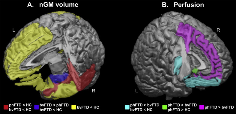
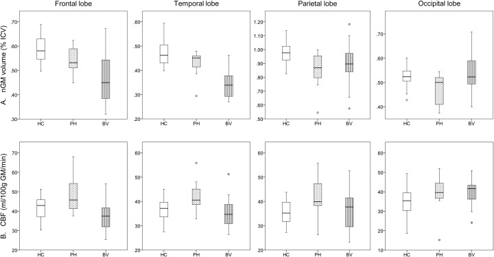
PhFTD patients showed cortical atrophy, most prominently in the right temporal lobe. Apart from this regional atrophy, GM volume was generally not different from either controls or from bvFTD. BvFTD however showed extensive frontotemporal atrophy. Perfusion was increased in the left prefrontal cortex compared to bvFTD and to a lesser extent to controls.
PhFTD and bvFTD show overlapping cortical structural abnormalities indicating a continuum of changes especially in the frontotemporal regions. Together with functional changes suggestive of a compensatory response to incipient pathology in the left prefrontal regions, these findings are the first to support a possible neuropathological etiology of phFTD and suggest that phFTD may be a neurodegenerative disease on the FTD spectrum.
“拟表型”额颞叶痴呆(phFTD)患者在临床上可能类似于额颞叶痴呆的行为变异型(bvFTD),但在常规神经影像学的视觉检查中未显示功能下降或异常。我们旨在识别phFTD患者灰质(GM)体积和灌注的异常,并评估phFTD是否属于额颞叶痴呆谱系。我们将phFTD患者与健康对照者和bvFTD患者进行了比较。
7例phFTD患者、11例bvFTD患者以及20例年龄匹配的对照者在3T条件下接受了结构T1加权磁共振成像(MRI)和三维伪连续动脉自旋标记(pCASL)检查。对经部分容积效应校正后的标准化GM(nGM)体积和灌注进行了区域以及整个幕上皮质的定量分析,并在考虑性别和扫描仪潜在混杂效应的情况下对组间进行了比较。
phFTD患者表现出皮质萎缩,最明显的是在右侧颞叶。除了这种局部萎缩外,GM体积总体上与对照组或bvFTD组没有差异。然而,bvFTD表现出广泛的额颞叶萎缩。与bvFTD相比,左侧前额叶皮质的灌注增加,与对照组相比增加程度较小。
phFTD和bvFTD表现出重叠的皮质结构异常,表明存在连续的变化,尤其是在额颞叶区域。这些发现连同提示对左侧前额叶区域早期病理变化有代偿反应的功能变化,首次支持了phFTD可能的神经病理学病因,并提示phFTD可能是额颞叶痴呆谱系中的一种神经退行性疾病。