Qin Chaoyi, Zhang Hongwei, Gu Jun, Xiao Zhenghua, Yang Qin, Meng Wei
Department of Cardiovascular Surgery, West China Hospital, Sichuan University, Lane outside the southern No. 37, Cheng du, Sichuan, People's Republic of China.
Department of Radiology, West China Hospital, Sichuan University, Lane outside the southern No. 37, Cheng du, Sichuan, People's Republic of China.
J Cardiothorac Surg. 2016 May 26;11(1):86. doi: 10.1186/s13019-016-0472-5.
To confirm the activation of platelets (PLT) and explore the role of activated PLT in post-dissection inflammation.
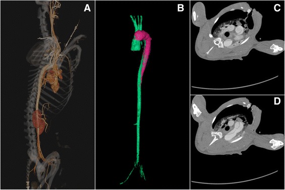
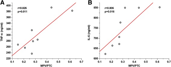
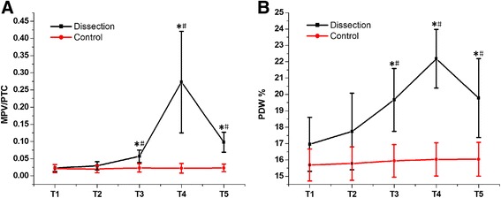
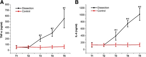
An acute type A aortic dissection (AAD) canine model was established. Mean platelet volume/platelet count (MPV/PTC), platelet size distribution width (PDW), and inflammatory cytokines (tumor necrosis factor-α [TNF-α] and interleukin-6 [IL-6]) were measured between anesthetization and thoracotomy (T1), at the end of the operation (T2), and at 2 h (T3), 4 h (T4), and 6 h (T5) after the operation. Bivariate analysis was used to determine the correlations between the peak MPV/PTC, PDW, and inflammatory cytokines at T4.
An AAD canine model was successfully established. Both MPV/PTC and PDW values were significantly higher at T3-T5 than at T1 (P < 0.05). Both were also significantly higher at T3-T5 in the dissection group than in the sham operation (SO) group (P < 0.05). Inflammatory cytokine levels were remarkably higher at T3-T5 than at T1, and were higher at T3-T5 in both the dissection and the SO group (P < 0.05). Bivariate analysis demonstrated positive correlations between MPV/PTC and both TNF-α (r = 0.826, P = 0.011) and IL-6 (r = 0.806, P = 0.016).
Activated PLT were identified after AAD, and played a critical role in the initiation of post-dissection inflammation.
确认血小板(PLT)的激活,并探讨活化血小板在夹层分离后炎症中的作用。
建立急性A型主动脉夹层(AAD)犬模型。在麻醉与开胸手术之间(T1)、手术结束时(T2)以及术后2小时(T3)、4小时(T4)和6小时(T5)测量平均血小板体积/血小板计数(MPV/PTC)、血小板体积分布宽度(PDW)和炎性细胞因子(肿瘤坏死因子-α [TNF-α]和白细胞介素-6 [IL-6])。采用双变量分析确定T4时MPV/PTC、PDW峰值与炎性细胞因子之间的相关性。
成功建立AAD犬模型。T3 - T5时MPV/PTC和PDW值均显著高于T1(P < 0.05)。夹层分离组T3 - T5时两者也显著高于假手术(SO)组(P < 0.05)。T3 - T5时炎性细胞因子水平显著高于T1,夹层分离组和SO组T3 - T5时均较高(P < 0.05)。双变量分析显示MPV/PTC与TNF-α(r = 0.826,P = 0.011)和IL-6(r = 0.806,P = 0.016)均呈正相关。
AAD后可识别出活化血小板,其在夹层分离后炎症的起始中起关键作用。