Mortensen Lena S, Park Silvia J H, Ke Jiang-Bin, Cooper Benjamin H, Zhang Lei, Imig Cordelia, Löwel Siegrid, Reim Kerstin, Brose Nils, Demb Jonathan B, Rhee Jeong-Seop, Singer Joshua H
Department of Molecular Neurobiology, Max Planck Institute of Experimental Medicine, 37075 Göttingen, Germany.
Department of Ophthalmology and Visual Science, Yale University, New Haven, CT 06511, USA.
Cell Rep. 2016 Jun 7;15(10):2239-2250. doi: 10.1016/j.celrep.2016.05.012. Epub 2016 May 26.
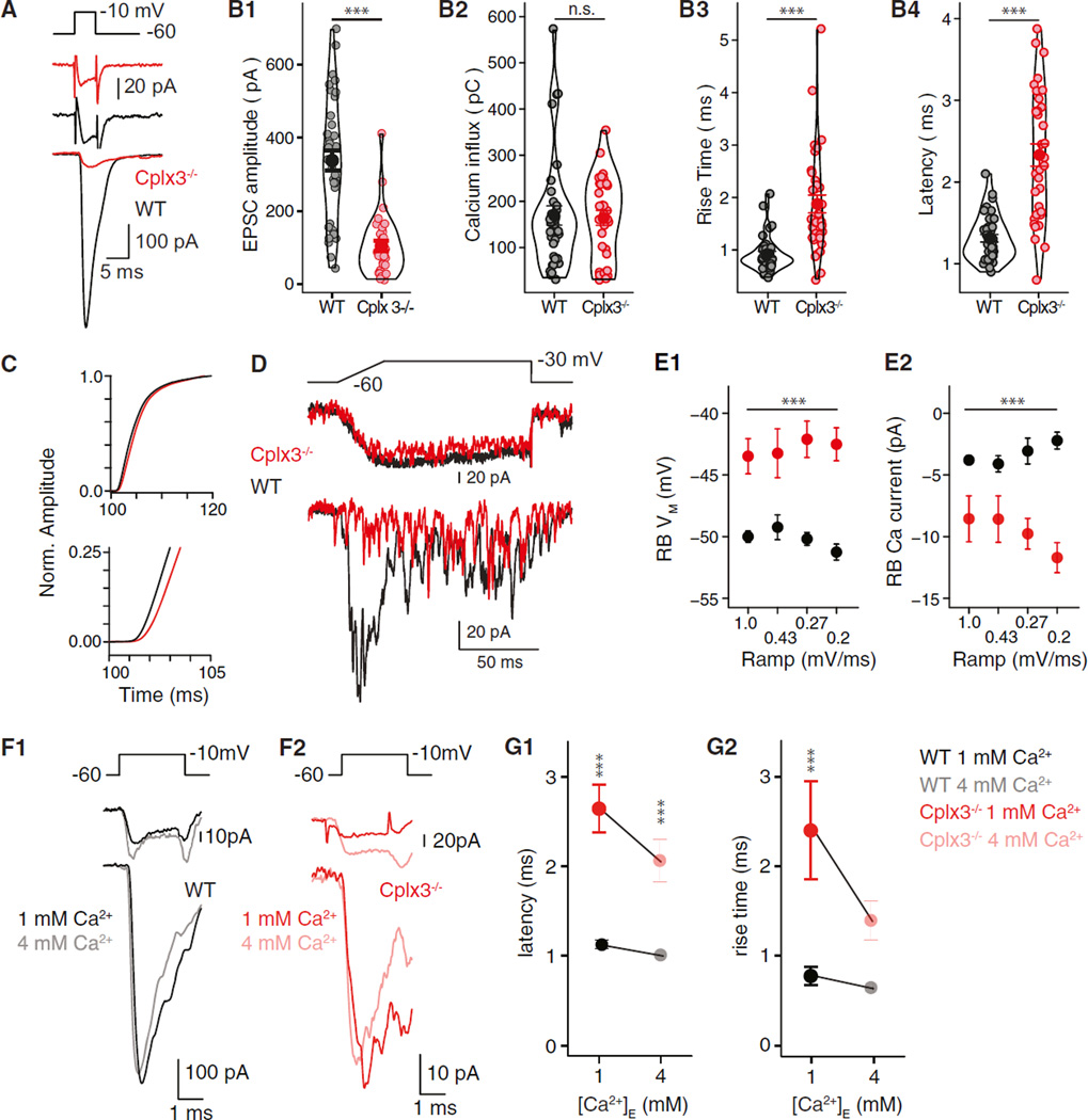
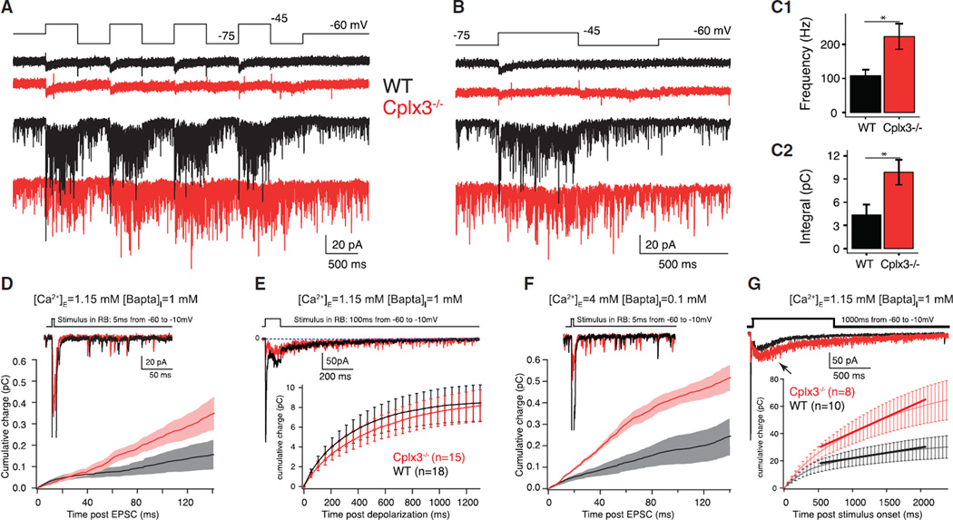
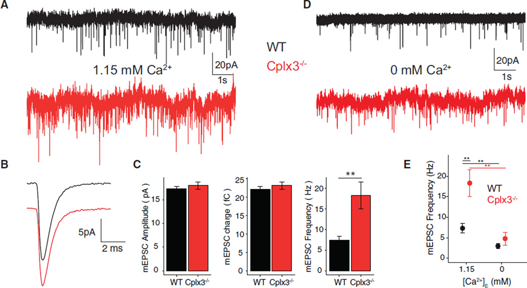
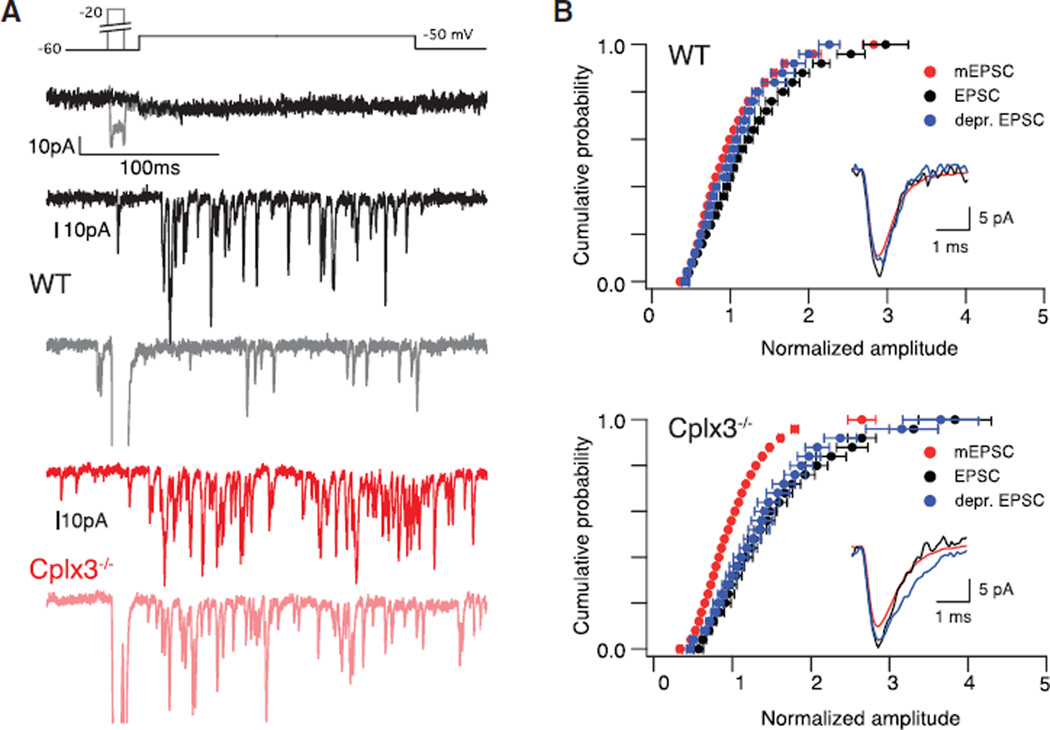
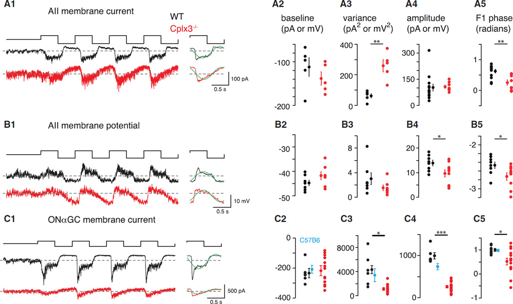
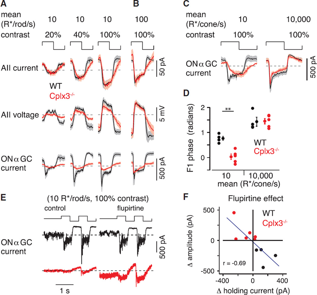
Complexin (Cplx) proteins modulate the core SNARE complex to regulate exocytosis. To understand the contributions of Cplx to signaling in a well-characterized neural circuit, we investigated how Cplx3, a retina-specific paralog, shapes transmission at rod bipolar (RB)→AII amacrine cell synapses in the mouse retina. Knockout of Cplx3 strongly attenuated fast, phasic Ca(2+)-dependent transmission, dependent on local [Ca(2+)] nanodomains, but enhanced slower Ca(2+)-dependent transmission, dependent on global intraterminal [Ca(2+)] ([Ca(2+)]I). Surprisingly, coordinated multivesicular release persisted at Cplx3(-/-) synapses, although its onset was slowed. Light-dependent signaling at Cplx3(-/-) RB→AII synapses was sluggish, owing largely to increased asynchronous release at light offset. Consequently, propagation of RB output to retinal ganglion cells was suppressed dramatically. Our study links Cplx3 expression with synapse and circuit function in a specific retinal pathway and reveals a role for asynchronous release in circuit gain control.
复合体(Cplx)蛋白调节核心SNARE复合体以调控胞吐作用。为了解Cplx在一个特征明确的神经回路中对信号传导的作用,我们研究了视网膜特异性旁系同源物Cplx3如何塑造小鼠视网膜中视杆双极(RB)细胞→无长突AII细胞突触处的传递。敲除Cplx3强烈减弱了快速、相位性钙(Ca2+)依赖性传递,该传递依赖于局部[Ca2+]纳米域,但增强了较慢的钙(Ca2+)依赖性传递,该传递依赖于终末内整体[Ca2+]([Ca2+]I)。令人惊讶的是,尽管其起始变慢,但在Cplx3基因敲除的突触处,协同多泡释放仍持续存在。Cplx3基因敲除的RB→AII突触处的光依赖性信号传导迟缓,这主要是由于在光熄灭时异步释放增加所致。因此,RB输出向视网膜神经节细胞的传播被显著抑制。我们的研究将Cplx3的表达与特定视网膜通路中的突触和回路功能联系起来,并揭示了异步释放在回路增益控制中的作用。