Zikopoulos Basilis, John Yohan J, García-Cabezas Miguel Ángel, Bunce Jamie G, Barbas Helen
Human Systems Neuroscience Laboratory, Department of Health Sciences, Boston University, Boston, MA, United States; Graduate Program for Neuroscience, Boston University and School of Medicine, Boston, MA, United States.
Neural Systems Laboratory, Department of Health Sciences, Boston University, Boston, MA, United States.
Neuroscience. 2016 Aug 25;330:267-90. doi: 10.1016/j.neuroscience.2016.05.052. Epub 2016 May 30.
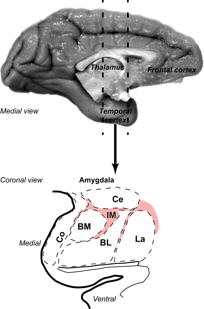
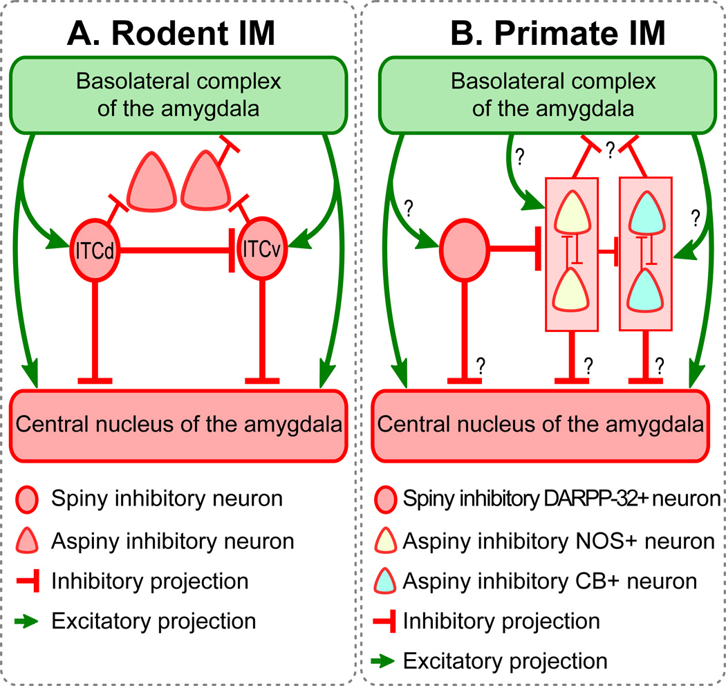
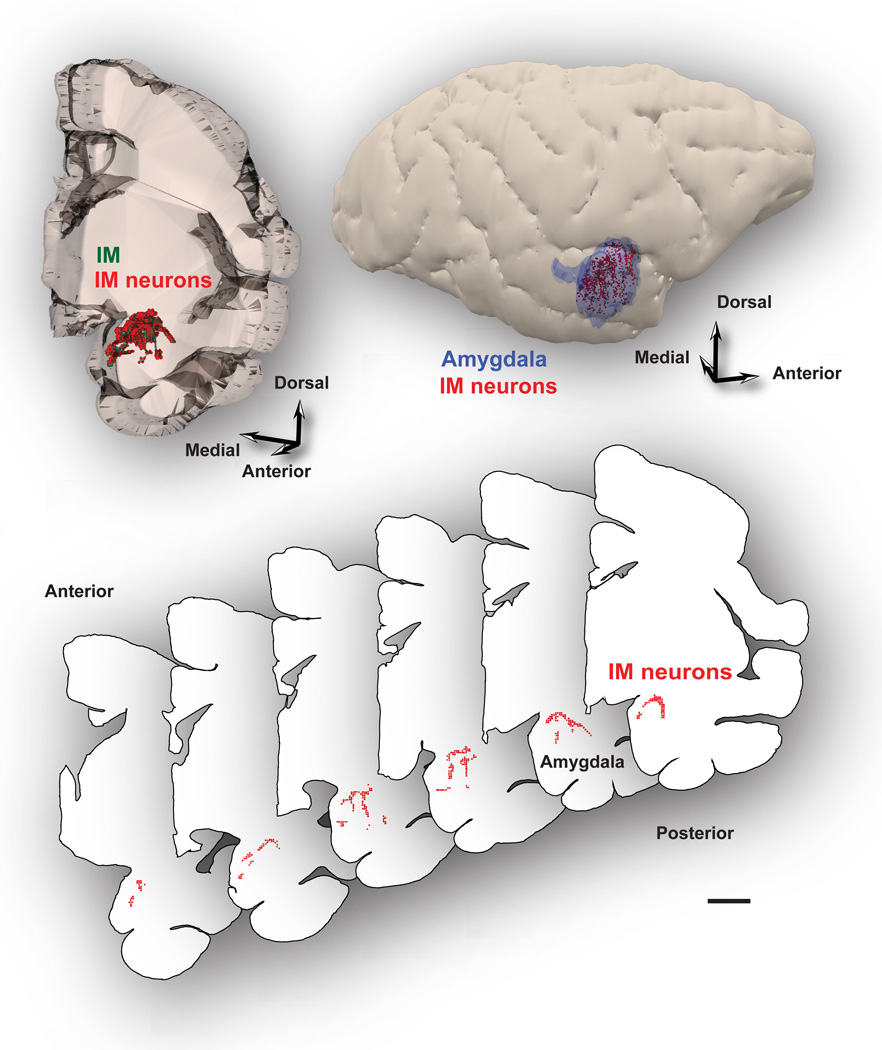
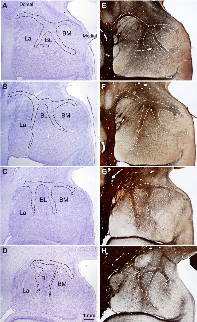
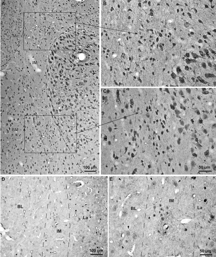
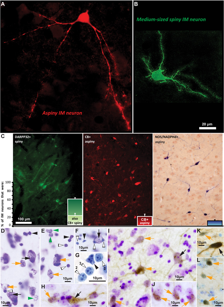
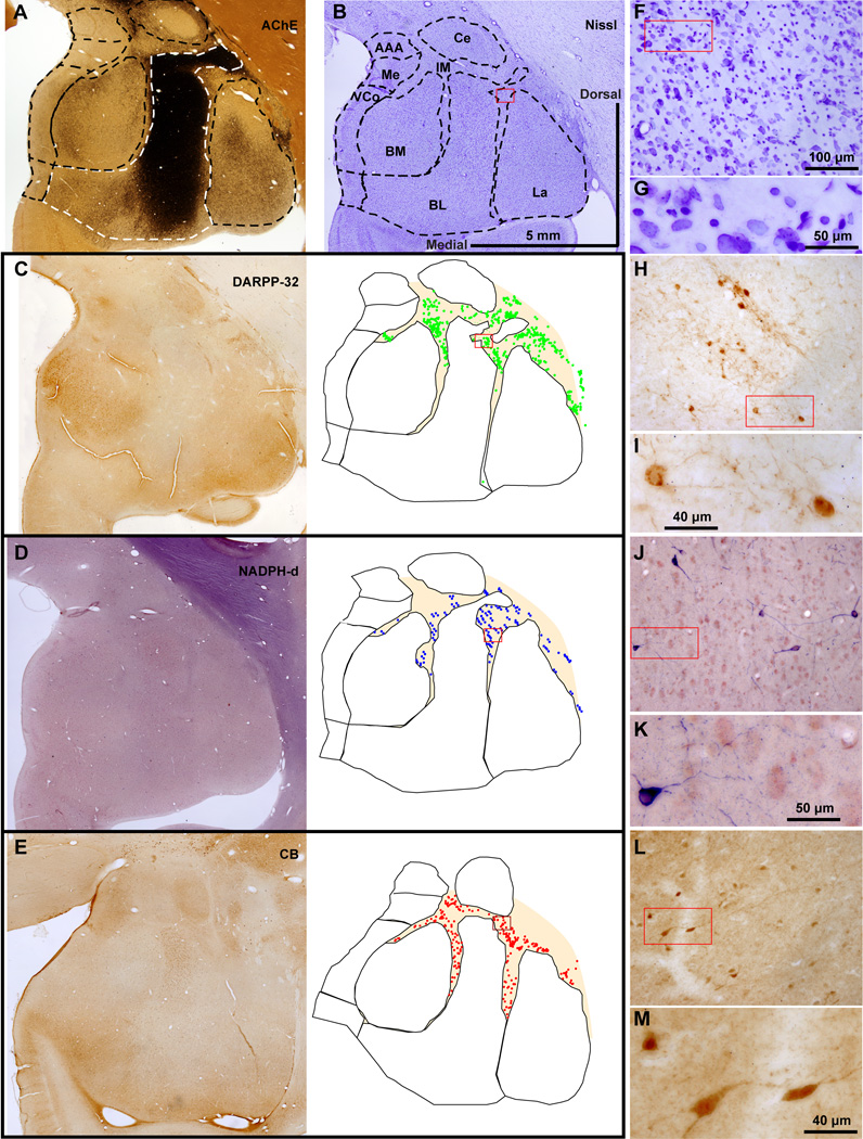
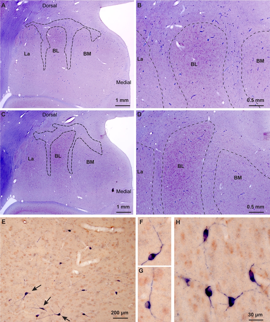
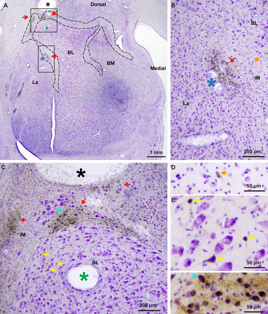
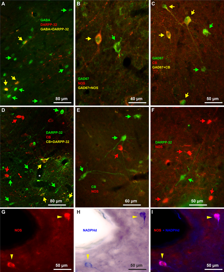
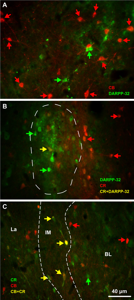
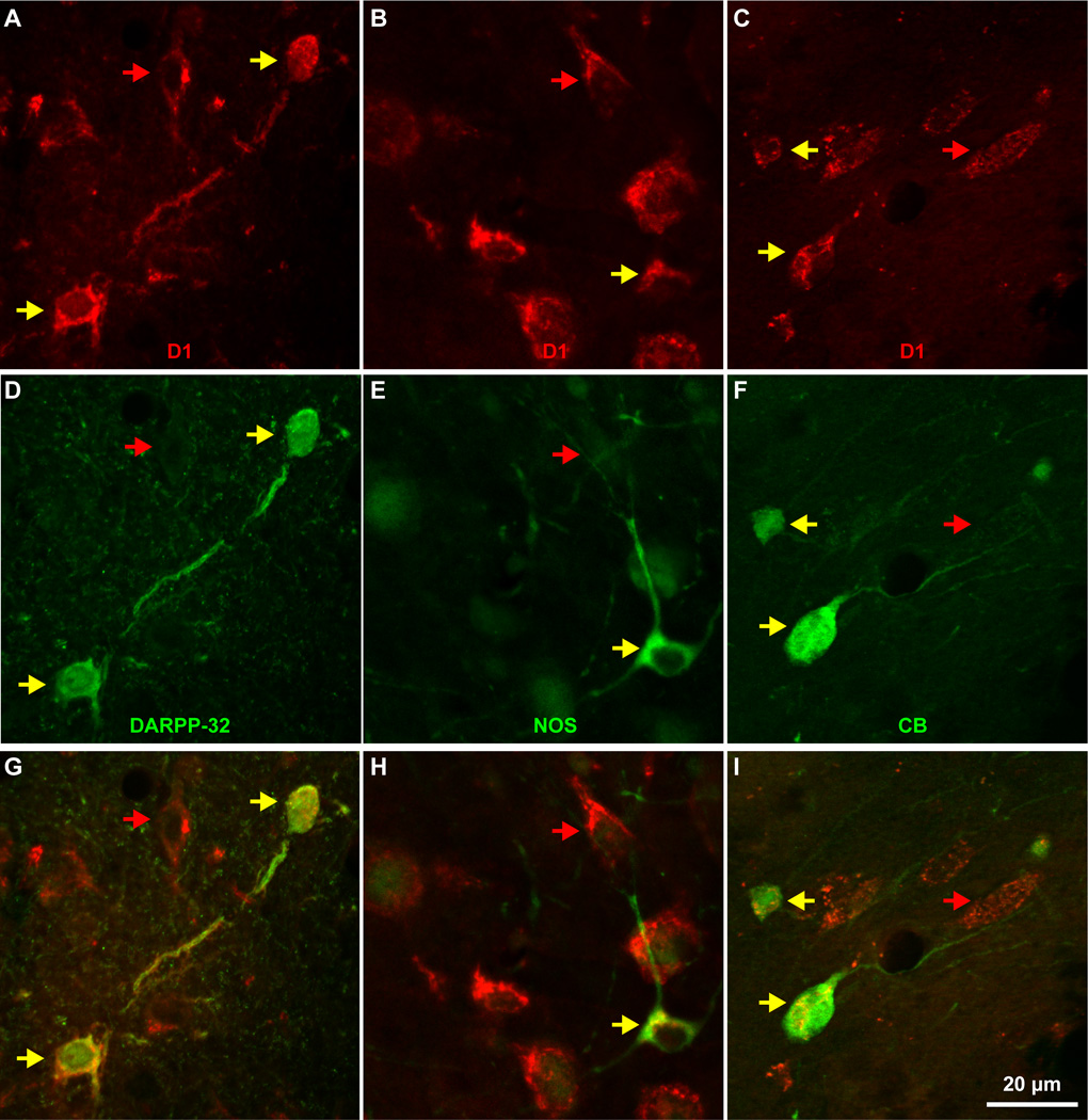
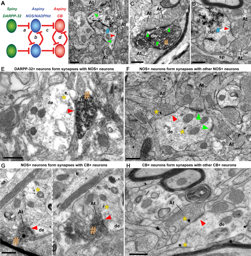
The organization of the inhibitory intercalated cell masses (IM) of the primate amygdala is largely unknown despite their key role in emotional processes. We studied the structural, topographic, neurochemical and intrinsic connectional features of IM neurons in the rhesus monkey brain. We found that the intercalated neurons are not confined to discrete cell clusters, but form a neuronal net that is interposed between the basal nuclei and extends to the dorsally located anterior, central, and medial nuclei of the amygdala. Unlike the IM in rodents, which are prominent in the anterior half of the amygdala, the primate inhibitory net stretched throughout the antero-posterior axis of the amygdala, and was most prominent in the central and posterior extent of the amygdala. There were two morphologic types of intercalated neurons: spiny and aspiny. Spiny neurons were the most abundant; their somata were small or medium size, round or elongated, and their dendritic trees were round or bipolar, depending on location. The aspiny neurons were on average slightly larger and had varicose dendrites with no spines. There were three non-overlapping neurochemical populations of IM neurons, in descending order of abundance: (1) Spiny neurons that were positive for the striatal associated dopamine- and cAMP-regulated phosphoprotein (DARPP-32+); (2) Aspiny neurons that expressed the calcium-binding protein calbindin (CB+); and (3) Aspiny neurons that expressed nitric oxide synthase (NOS+). The unique combinations of structural and neurochemical features of the three classes of IM neurons suggest different physiological properties and function. The three types of IM neurons were intermingled and likely interconnected in distinct ways, and were innervated by intrinsic neurons within the amygdala, or by external sources, in pathways that underlie fear conditioning and anxiety.
尽管抑制性闰细胞团(IM)在灵长类动物杏仁核的情感过程中起着关键作用,但其组织结构在很大程度上仍不为人所知。我们研究了恒河猴大脑中IM神经元的结构、拓扑、神经化学和内在连接特征。我们发现,闰细胞并不局限于离散的细胞簇,而是形成一个神经元网络,该网络介于基底核之间,并延伸至杏仁核背侧的前核、中央核和内侧核。与啮齿动物的IM不同,后者在杏仁核的前半部分较为突出,而灵长类动物的抑制性网络贯穿杏仁核的前后轴,在杏仁核的中央和后部最为突出。闰细胞有两种形态类型:有棘和无棘。有棘神经元最为丰富;它们的胞体小或中等大小,呈圆形或椭圆形,其树突根据位置呈圆形或双极形。无棘神经元平均略大,有曲张的树突且无棘。IM神经元有三个不重叠的神经化学群体,按丰度降序排列:(1)对纹状体相关多巴胺和cAMP调节磷蛋白呈阳性的有棘神经元(DARPP - 32+);(2)表达钙结合蛋白钙结合蛋白的无棘神经元(CB+);(3)表达一氧化氮合酶的无棘神经元(NOS+)。这三类IM神经元独特的结构和神经化学特征组合表明它们具有不同的生理特性和功能。这三种类型的IM神经元相互交织,并可能以不同的方式相互连接,并且在恐惧条件反射和焦虑所基于的通路中,由杏仁核内的内在神经元或外部来源支配。