Niranjan Arun, Christie Isabel N, Solomon Samuel G, Wells Jack A, Lythgoe Mark F
UCL Centre for Advanced Biomedical Imaging, Division of Medicine and Institute of Child Health, University College London, London, UK.
UCL Centre for Advanced Biomedical Imaging, Division of Medicine and Institute of Child Health, University College London, London, UK; Centre for Cardiovascular and Metabolic Neuroscience, Department of Neuroscience, Physiology & Pharmacology, University College London, London, UK.
Neuroimage. 2016 Oct 1;139:337-345. doi: 10.1016/j.neuroimage.2016.06.015. Epub 2016 Jun 10.
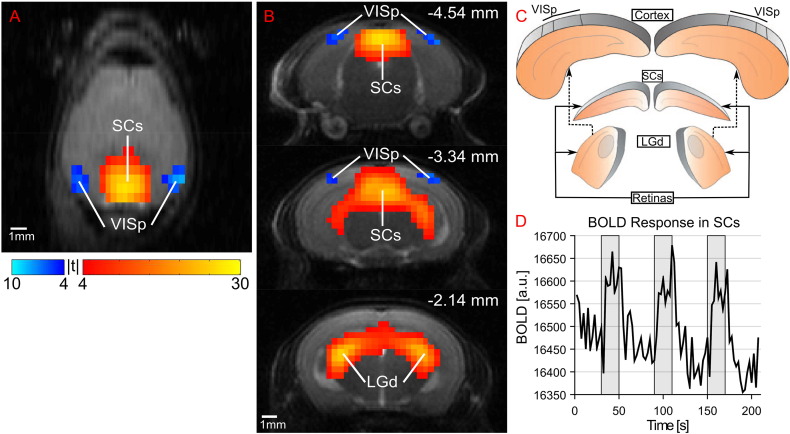
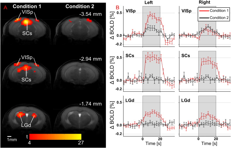
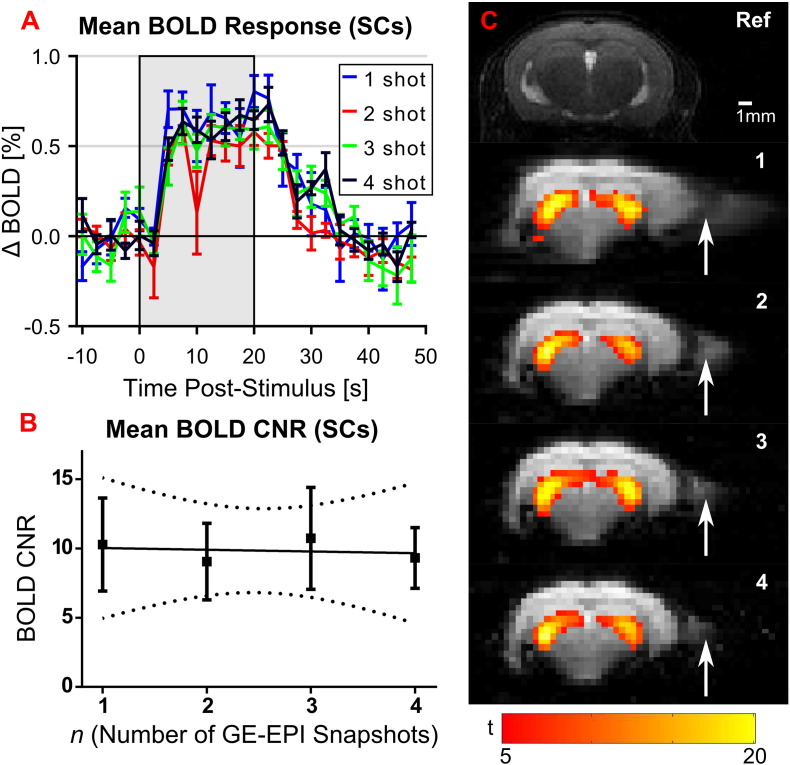
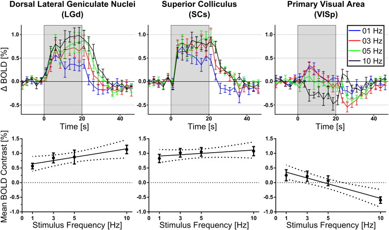
The use of functional magnetic resonance imaging (fMRI) in mice is increasingly prevalent, providing a means to non-invasively characterise functional abnormalities associated with genetic models of human diseases. The predominant stimulus used in task-based fMRI in the mouse is electrical stimulation of the paw. Task-based fMRI in mice using visual stimuli remains underexplored, despite visual stimuli being common in human fMRI studies. In this study, we map the mouse brain visual system with BOLD measurements at 9.4T using flashing light stimuli with medetomidine anaesthesia. BOLD responses were observed in the lateral geniculate nucleus, the superior colliculus and the primary visual area of the cortex, and were modulated by the flashing frequency, diffuse vs focussed light and stimulus context. Negative BOLD responses were measured in the visual cortex at 10Hz flashing frequency; but turned positive below 5Hz. In addition, the use of interleaved snapshot GE-EPI improved fMRI image quality without diminishing the temporal contrast-noise-ratio. Taken together, this work demonstrates a novel methodological protocol in which the mouse brain visual system can be non-invasively investigated using BOLD fMRI.
功能磁共振成像(fMRI)在小鼠中的应用越来越普遍,它为非侵入性地表征与人类疾病遗传模型相关的功能异常提供了一种手段。小鼠基于任务的fMRI中使用的主要刺激是对爪子进行电刺激。尽管视觉刺激在人类fMRI研究中很常见,但在小鼠中使用视觉刺激进行基于任务的fMRI仍未得到充分探索。在本研究中,我们在9.4T磁场下,使用美托咪定麻醉并施加闪光刺激,通过血氧水平依赖(BOLD)测量来绘制小鼠脑视觉系统图谱。在外侧膝状体、上丘和皮层的初级视觉区观察到了BOLD反应,并且这些反应受到闪光频率、漫射光与聚焦光以及刺激背景的调制。在10Hz闪光频率下,视觉皮层中测量到负性BOLD反应;但在5Hz以下变为正性。此外,使用交错快照梯度回波平面成像(GE-EPI)可提高fMRI图像质量,同时不降低时间对比度噪声比。综上所述,这项工作展示了一种新颖的方法学方案,即可以使用BOLD fMRI对小鼠脑视觉系统进行非侵入性研究。