State Key Laboratory of Respiratory Disease, National Clinical Research Center for Respiratory Disease, Guangzhou Institute of Respiratory Disease, First Affiliated Hospital of Guangzhou Medical University, Guangzhou, Guangdong, China.
Department of Respiratory and Critical Care Medicine, First Affiliated Hospital of Zhengzhou University, Zhengzhou, Henan, China.
Sci Rep. 2016 Jun 24;6:28467. doi: 10.1038/srep28467.
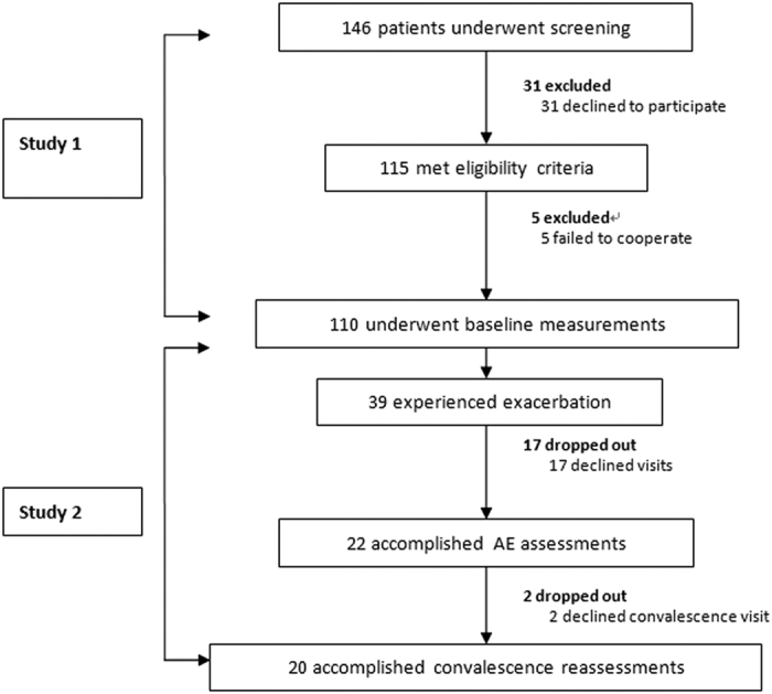
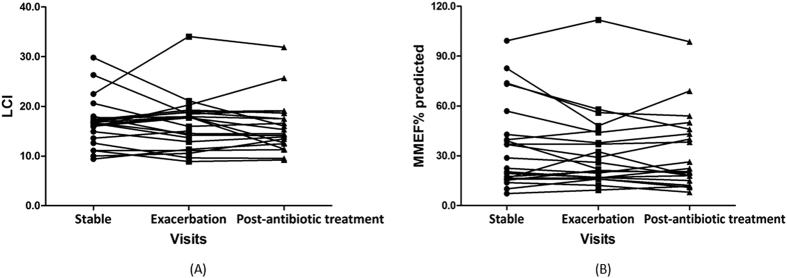
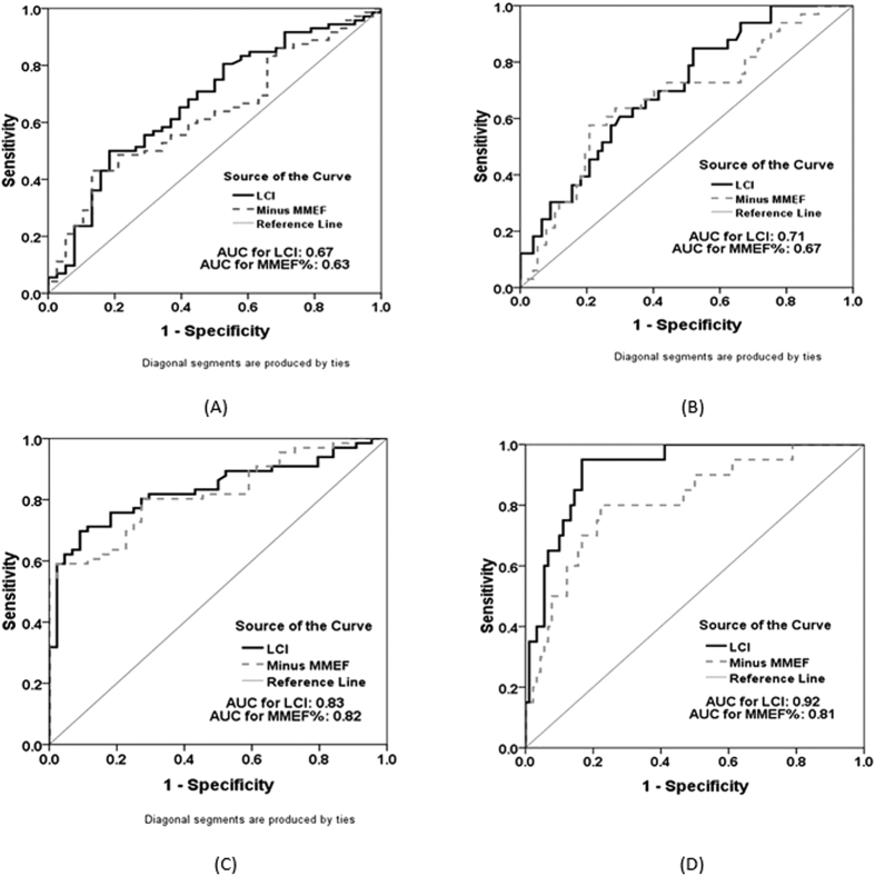
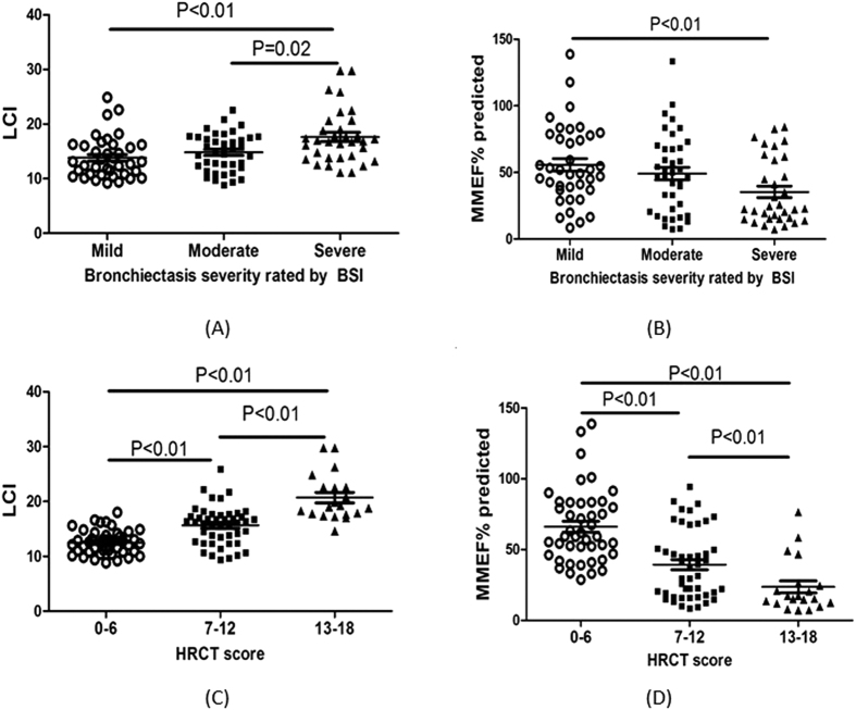
Little is known about the comparative diagnostic value of lung clearance index (LCI) and maximal mid-expiratory flow (MMEF) in bronchiectasis. We compared the diagnostic performance, correlation and concordance with clinical variables, and changes of LCI and MMEF% predicted during bronchiectasis exacerbations (BEs). Patients with stable bronchiectasis underwent history inquiry, chest high-resolution computed tomography (HRCT), multiple-breath nitrogen wash-out test, spirometry and sputum culture. Patients who experienced BEs underwent these measurements during onset of BEs and 1 week following antibiotics therapy. Sensitivity analyses were performed in mild, moderate and severe bronchiectasis. We recruited 110 bronchiectasis patients between March 2014 and September 2015. LCI demonstrated similar diagnostic value with MMEF% predicted in discriminating moderate-to-severe from mild bronchiectasis. LCI negatively correlated with MMEF% predicted. Both parameters had similar concordance in reflecting clinical characteristics of bronchiectasis and correlated significantly with forced expiratory flow in one second, age, HRCT score, Pseudomonas aeruginosa colonization, cystic bronchiectasis, ventilation heterogeneity and bilateral bronchiectasis. In exacerbation cohort (n = 22), changes in LCI and MMEF% predicted were equally minimal during BEs and following antibiotics therapy. In sensitivity analyses, both parameters had similar diagnostic value and correlation with clinical variables. MMEF% predicted is a surrogate of LCI for assessing bronchiectasis severity.
关于肺清除指数 (LCI) 和最大呼气中期流量 (MMEF) 在支气管扩张症中的比较诊断价值知之甚少。我们比较了 LCI 和 MMEF%预测值在支气管扩张症加重 (BE) 期间的诊断性能、相关性和与临床变量的一致性,以及变化。稳定期支气管扩张症患者接受病史询问、胸部高分辨率计算机断层扫描 (HRCT)、多次呼吸氮冲洗试验、肺活量测定和痰培养。经历 BE 的患者在 BE 发作时和抗生素治疗后 1 周进行这些测量。在轻度、中度和重度支气管扩张症中进行了敏感性分析。我们于 2014 年 3 月至 2015 年 9 月期间招募了 110 名支气管扩张症患者。LCI 在区分中重度和轻度支气管扩张症方面与 MMEF%预测值具有相似的诊断价值。LCI 与 MMEF%预测值呈负相关。这两个参数在反映支气管扩张症的临床特征方面具有相似的一致性,并且与用力呼气第一秒、年龄、HRCT 评分、铜绿假单胞菌定植、囊状支气管扩张、通气异质性和双侧支气管扩张显著相关。在加重组(n=22)中,在 BE 期间和抗生素治疗后,LCI 和 MMEF%预测值的变化同样微小。在敏感性分析中,这两个参数在诊断价值和与临床变量的相关性方面具有相似性。MMEF%预测值是评估支气管扩张症严重程度的 LCI 替代指标。