Said Elias A, Al-Reesi Iman, Al-Riyami Marwa, Al-Naamani Khalid, Al-Sinawi Shadia, Al-Balushi Mohammed S, Koh Crystal Y, Al-Busaidi Juma Z, Idris Mohamed A, Al-Jabri Ali A
Department of Microbiology and Immunology, College of Medicine and Health Sciences, Sultan Qaboos University, P.O. Box: 35, Code: 123, Muscat, Oman.
Department of Pathology, College of Medicine and Health Sciences, Sultan Qaboos University, P.O. Box: 35, Code: 123, Muscat, Oman.
PLoS One. 2016 Jun 27;11(6):e0158265. doi: 10.1371/journal.pone.0158265. eCollection 2016.
The failure to establish potent anti-HBV T cell responses suggests the absence of an effective innate immune activation. Kupffer cells and liver-infiltrating monocytes/macrophages have an essential role in establishing anti-HBV responses. These cells express the costimulatory molecules CD80 and CD86. CD80 expression on antigen-presenting cells (APCs) induces Th1 cell differentiation, whereas CD86 expression drives the differentiation towards a Th2 profile. The relative expression of CD80, CD86 and PD-L1 on APCs, regulates T cell activation. Few studies investigated CD80 and CD86 expression on KCs and infiltrating monocytes/macrophages in HBV-infected liver and knowledge about the expression of PD-L1 on these cells is controversial. The expression of these molecules together in CD68+ cells has not been explored in HBV-infected livers.
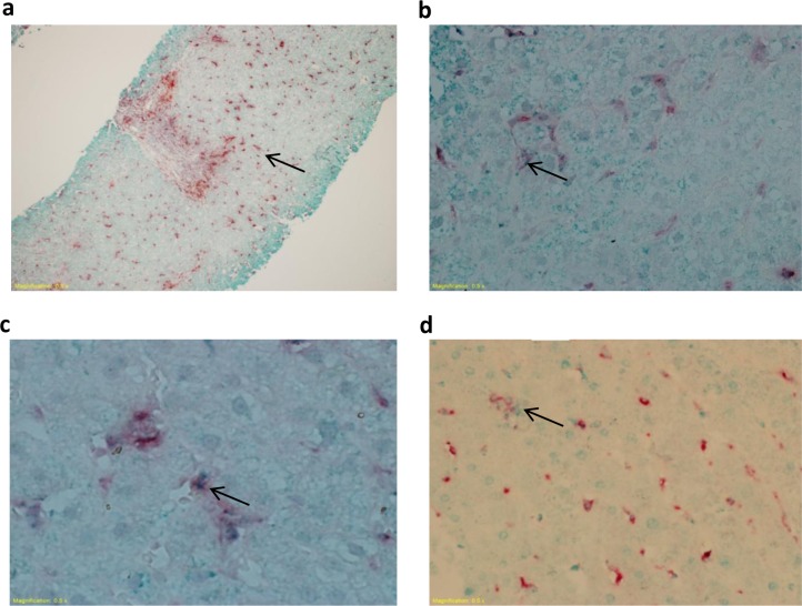
Double staining immunohistochemistry was applied to liver biopsies of HBV-infected and control donors to explore CD80, CD86 and PD-L1 expression in the lobular and portal areas.
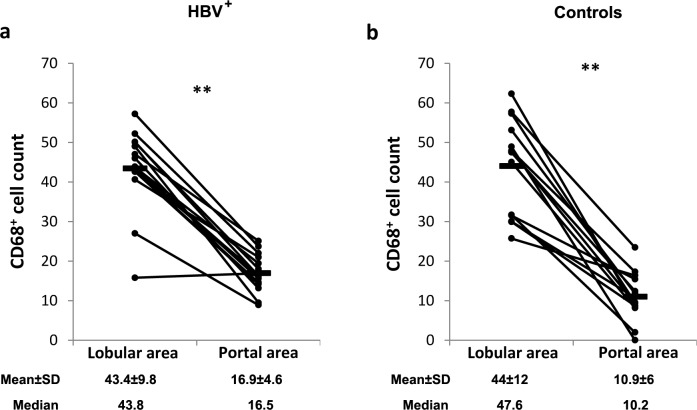
Chronic HBV infection was associated with increased CD68+CD86+ cell count and percentage in the lobular areas, and no changes in the count and percentage of CD68+CD80+ and CD68+PD-L1+ cells, compared to the control group. While CD68+CD80+ cell count in portal areas correlated with the fibrosis score, CD68+CD80+ cell percentage in lobular areas correlated with the inflammation grade.
The upregulation of CD86 but not CD80 and PD-L1 on CD68+ cells in HBV-infected livers, suggests that these cells do not support the induction of potent Th1. Moreover, the expression of CD80 on CD68+ cells correlates with liver inflammation and fibrosis.
未能建立有效的抗乙肝病毒T细胞反应提示缺乏有效的天然免疫激活。库普弗细胞和肝内浸润的单核细胞/巨噬细胞在建立抗乙肝病毒反应中起重要作用。这些细胞表达共刺激分子CD80和CD86。抗原呈递细胞(APC)上的CD80表达诱导Th1细胞分化,而CD86表达驱动向Th2型分化。APC上CD80、CD86和PD-L1的相对表达调节T细胞活化。很少有研究调查乙肝病毒感染肝脏中库普弗细胞和浸润单核细胞/巨噬细胞上CD80和CD86的表达,关于这些细胞上PD-L1表达的知识存在争议。在乙肝病毒感染的肝脏中,尚未探讨这些分子在CD68+细胞中的共同表达情况。
对乙肝病毒感染和对照供体的肝活检组织进行双重染色免疫组化,以探究小叶和门管区中CD80、CD86和PD-L1的表达。
与对照组相比,慢性乙肝病毒感染与小叶区CD68+CD86+细胞计数和百分比增加有关,而CD68+CD80+和CD68+PD-L1+细胞的计数和百分比无变化。虽然门管区CD68+CD80+细胞计数与纤维化评分相关,但小叶区CD68+CD80+细胞百分比与炎症分级相关。
乙肝病毒感染肝脏中CD68+细胞上CD86而非CD80和PD-L1的上调表明,这些细胞不支持强效Th1的诱导。此外,CD68+细胞上CD80的表达与肝脏炎症和纤维化相关。