Zhang Ying, Cai Ai-Lu, Ren Wei-Dong, Guo Ya-Jun, Zhang Dong-Yu, Sun Wei, Wang Yu, Wang Lei, Qin Yue, Huang Li-Ping
Department of Sonography, Shengjing Hospital of China Medical University, No. 36 Sanhao Street, Heping District, Shenyang, Liaoning, China.
BMC Pregnancy Childbirth. 2016 Jun 30;16:145. doi: 10.1186/s12884-016-0933-9.
Prenatal cardiac screening is of great importance as it contributes to appropriate neonatal management and helps parents to make a decision regarding their pregnancy. The aim of our study was to evaluate the efficiency of a newly proposed screening protocol in the detection of fetal congenital heart disease (CHD).
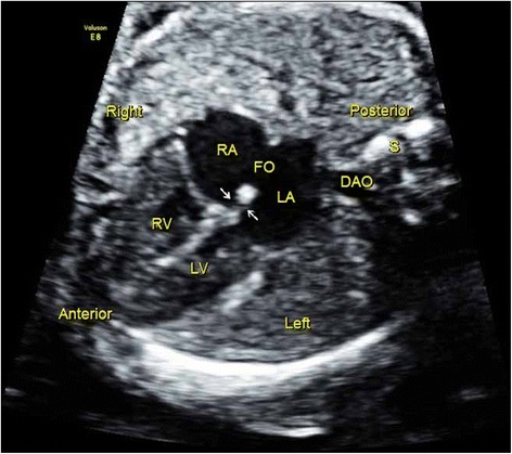
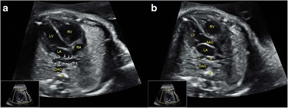
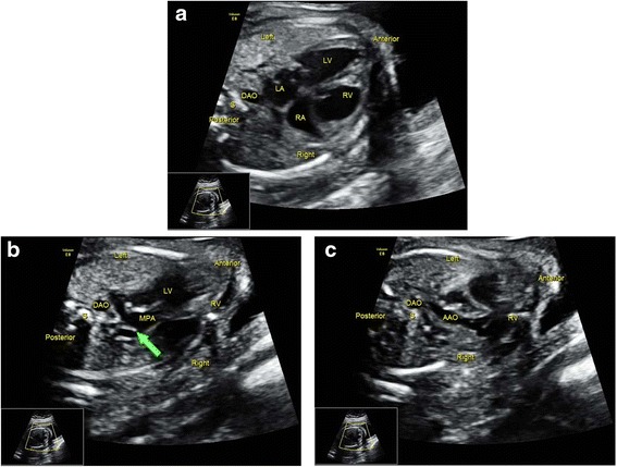
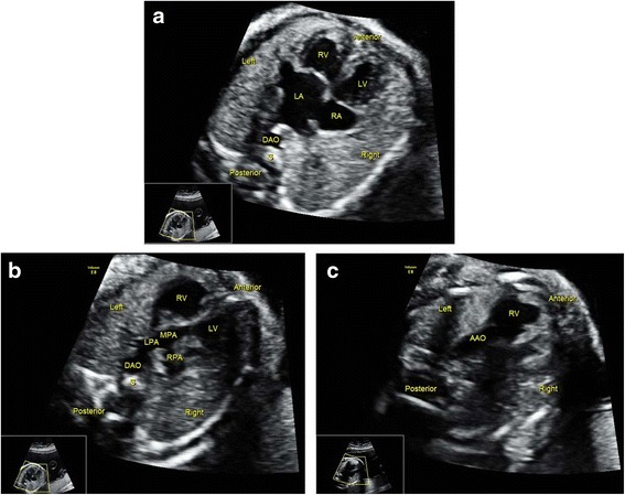
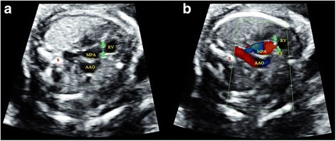
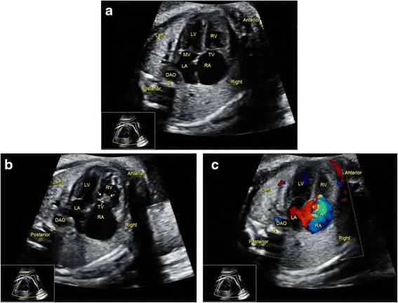
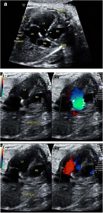
This was a prospective study. A total of 52 cases of confirmed CHD fetuses and 248 cases of randomly selected normal fetuses were included in the study. Two sonographers with similar experience performed the cardiac screenings under two different protocols independently. The conventional protocol (Protocol A) paid greater attention to the four-chamber view and the outflow tract views. A 6-month training program was provided to sonographers performing scans under the new protocol (Protocol B), which emphasized systematically evaluating fetal cardiac anatomy and hemodynamics. Color Doppler was mandatory and some ultrasonic signs for special cardiac anomalies were also introduced into this protocol.
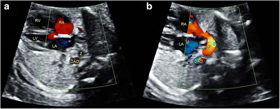
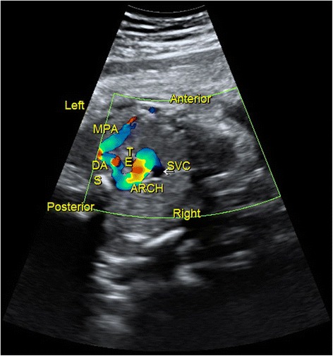
Protocol B detected more cardiac anomalies than did Protocol A (96.2 % vs. 61.5 %, P < 0.01). Specifically, Protocol B was superior to Protocol A in detecting cardiac malpositions, abnormal systemic and pulmonary venous connection, right aortic arch, transposition of the great arteries, and congenital corrected transposition of the great arteries. By visualizing flow disturbance and retrograde flow with color Doppler, Protocol B was better than Protocol A in screening valvular associated malformations, such as pulmonary atresia, pulmonary stenosis, tricuspid dysplasia, etc. For the normal fetuses, Protocol B was better than Protocol A in reducing the false-positive detection of septal defects.
The current study introduces an enhanced protocol for fetal cardiac screening, under which the obstetric screening sonographers systematically identify fetal cardiac anatomy and hemodynamics. A short-term training program makes it possible for the screening sonographers to become familiar with the new protocol, and its value has been confirmed due to improvements made in screening efficiency.
产前心脏筛查非常重要,因为它有助于新生儿的合理管理,并帮助父母就其妊娠做出决定。我们研究的目的是评估一种新提出的筛查方案在检测胎儿先天性心脏病(CHD)方面的效率。
这是一项前瞻性研究。研究共纳入52例确诊为CHD的胎儿和248例随机选择的正常胎儿。两名经验相似的超声检查医师分别按照两种不同的方案独立进行心脏筛查。传统方案(方案A)更注重四腔心切面和流出道切面。对按照新方案(方案B)进行扫描的超声检查医师提供了一个为期6个月的培训项目,该方案强调系统地评估胎儿心脏解剖结构和血流动力学。彩色多普勒检查是必需的,一些特殊心脏异常的超声征象也被纳入该方案。
方案B检测出的心脏异常比方案A多(96.2%对61.5%,P<0.01)。具体而言,方案B在检测心脏位置异常、体肺静脉连接异常、右位主动脉弓、大动脉转位和先天性矫正型大动脉转位方面优于方案A。通过彩色多普勒观察血流紊乱和逆流情况,方案B在筛查瓣膜相关畸形(如肺动脉闭锁、肺动脉狭窄、三尖瓣发育异常等)方面比方案A更好。对于正常胎儿,方案B在减少室间隔缺损的假阳性检测方面比方案A更好。
本研究引入了一种改进的胎儿心脏筛查方案,在该方案下,产科筛查超声检查医师可系统地识别胎儿心脏解剖结构和血流动力学。一个短期培训项目使筛查超声检查医师有可能熟悉新方案,并且由于筛查效率的提高,其价值已得到证实。