• 文献检索
  • 文档翻译
  • 深度研究
  • 学术资讯
  • Suppr Zotero 插件Zotero 插件
  • 邀请有礼
  • 套餐&价格
  • 历史记录
应用&插件
Suppr Zotero 插件Zotero 插件浏览器插件Mac 客户端Windows 客户端微信小程序
定价
高级版会员购买积分包购买API积分包
服务
文献检索文档翻译深度研究API 文档MCP 服务
关于我们
关于 Suppr公司介绍联系我们用户协议隐私条款
关注我们

Suppr 超能文献

核心技术专利:CN118964589B侵权必究
粤ICP备2023148730 号-1Suppr @ 2026

文献检索

告别复杂PubMed语法,用中文像聊天一样搜索,搜遍4000万医学文献。AI智能推荐,让科研检索更轻松。

立即免费搜索

文件翻译

保留排版,准确专业,支持PDF/Word/PPT等文件格式,支持 12+语言互译。

免费翻译文档

深度研究

AI帮你快速写综述,25分钟生成高质量综述,智能提取关键信息,辅助科研写作。

立即免费体验

Magnetic resonance imaging of spinal cord lesions in multiple sclerosis.

作者信息

Honig L S, Sheremata W A

机构信息

Department of Neurology, University of Miami School of Medicine, FL.

出版信息

J Neurol Neurosurg Psychiatry. 1989 Apr;52(4):459-66. doi: 10.1136/jnnp.52.4.459.

DOI:10.1136/jnnp.52.4.459
PMID:2738588
原文链接:https://pmc.ncbi.nlm.nih.gov/articles/PMC1032291/
Abstract

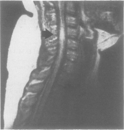
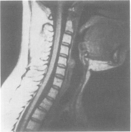
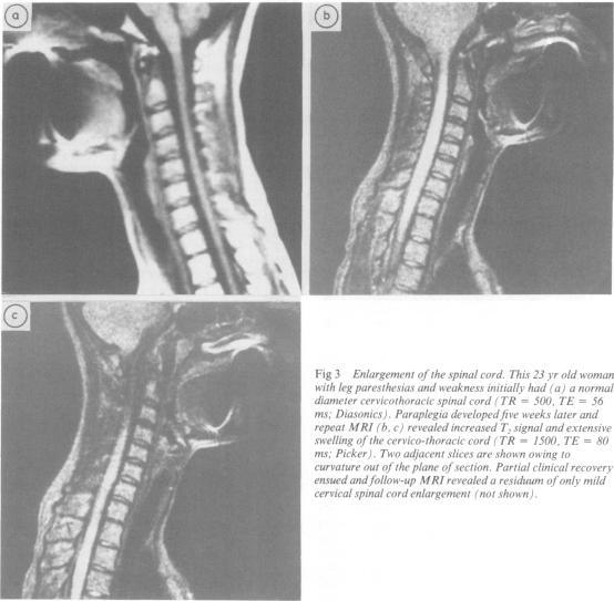
The clinical and pathological manifestations of multiple sclerosis are due to areas of demyelination which occur throughout the white matter of the central nervous system. MRI of the brain frequently shows abnormalities in the hemispheric subcortical white matter; these are demonstrable in the majority of patients and support the clinical diagnosis of multiple sclerosis. Our studies have shown that while MRI identifies such cerebral lesions in nearly all clinically definite multiple sclerosis patients with illness of duration greater than 10 years, these areas of abnormal T2 signal are present less often in the brains of patients studied within 3 years of disease onset. However, symptoms referable to the long tracts of the spinal cord are prominent in many of these patients. Imaging of the spinal cord has presented technical problems because of the small size of the cord, patient body, heart and respiratory movements, and limitations of surface coil technology. The spinal cord of 77 patients with multiple sclerosis have been imaged, revealing three types of abnormalities: (1) approximately half the cords show regions of abnormal T2 weighted signal; (2) during acute exacerbation, spinal cord enlargement (swelling) may be observed; (3) spinal cord atrophy (narrowing) is found particularly in patients with disease of longer duration and greater disability. Unlike the presence of brain lesions, the existence of spinal cord lesions of high T2 signal is not associated with increasing duration of disease but is correlated with disability status. Of patients with such lesions about one fifth did not exhibit brain lesions discernible by MRI.

摘要
https://cdn.ncbi.nlm.nih.gov/pmc/blobs/abc5/1032291/5f974cd0c66f/jnnpsyc00526-0034-a.jpg
https://cdn.ncbi.nlm.nih.gov/pmc/blobs/abc5/1032291/2865818371d0/jnnpsyc00526-0032-a.jpg
https://cdn.ncbi.nlm.nih.gov/pmc/blobs/abc5/1032291/54c7dfdece88/jnnpsyc00526-0033-a.jpg
https://cdn.ncbi.nlm.nih.gov/pmc/blobs/abc5/1032291/5f974cd0c66f/jnnpsyc00526-0034-a.jpg
https://cdn.ncbi.nlm.nih.gov/pmc/blobs/abc5/1032291/2865818371d0/jnnpsyc00526-0032-a.jpg
https://cdn.ncbi.nlm.nih.gov/pmc/blobs/abc5/1032291/54c7dfdece88/jnnpsyc00526-0033-a.jpg
https://cdn.ncbi.nlm.nih.gov/pmc/blobs/abc5/1032291/5f974cd0c66f/jnnpsyc00526-0034-a.jpg

相似文献

1
Magnetic resonance imaging of spinal cord lesions in multiple sclerosis.
J Neurol Neurosurg Psychiatry. 1989 Apr;52(4):459-66. doi: 10.1136/jnnp.52.4.459.
2
Evidence for early neurodegeneration in the cervical cord of patients with primary progressive multiple sclerosis.原发性进行性多发性硬化症患者颈髓早期神经退行性变的证据。
Brain. 2015 Jun;138(Pt 6):1568-82. doi: 10.1093/brain/awv086. Epub 2015 Apr 10.
3
Cortical neuronal densities and cerebral white matter demyelination in multiple sclerosis: a retrospective study.多发性硬化症中的皮质神经元密度和脑白质脱髓鞘:一项回顾性研究。
Lancet Neurol. 2018 Oct;17(10):870-884. doi: 10.1016/S1474-4422(18)30245-X. Epub 2018 Aug 22.
4
Brain and cord myelin water imaging: a progressive multiple sclerosis biomarker.脑和脊髓髓鞘水成像:一种进展性多发性硬化症生物标志物。
Neuroimage Clin. 2015 Oct 3;9:574-80. doi: 10.1016/j.nicl.2015.10.002. eCollection 2015.
5
Demyelinating lesions in the cervical cord in multiple sclerosis 10 years after onset of the disease. Correlation between MRI parameters and clinical course.多发性硬化症发病10年后颈髓的脱髓鞘病变。MRI参数与临床病程之间的相关性。
Neurol Neurochir Pol. 2007 May-Jun;41(3):229-33.
6
Three dimensional MRI estimates of brain and spinal cord atrophy in multiple sclerosis.多发性硬化症中脑和脊髓萎缩的三维磁共振成像估计
J Neurol Neurosurg Psychiatry. 1999 Mar;66(3):323-30. doi: 10.1136/jnnp.66.3.323.
7
Post-mortem high-resolution MRI of the spinal cord in multiple sclerosis: a correlative study with conventional MRI, histopathology and clinical phenotype.多发性硬化症脊髓的死后高分辨率磁共振成像:与传统磁共振成像、组织病理学及临床表型的相关性研究
Brain. 2001 Jan;124(Pt 1):154-66. doi: 10.1093/brain/124.1.154.
8
The role of NMR imaging in the assessment of multiple sclerosis and isolated neurological lesions. A quantitative study.核磁共振成像在多发性硬化症和孤立性神经病变评估中的作用。一项定量研究。
Brain. 1987 Dec;110 ( Pt 6):1579-616. doi: 10.1093/brain/110.6.1579.
9
Spinal cord magnetic resonance imaging in suspected multiple sclerosis.疑似多发性硬化症的脊髓磁共振成像
Eur Radiol. 2000;10(2):368-76. doi: 10.1007/s003300050058.
10
Mean upper cervical cord area (MUCCA) measurement in long-standing multiple sclerosis: relation to brain findings and clinical disability.长期多发性硬化症患者中上颈髓面积(MUCCA)的测量:与脑发现和临床残疾的关系。
Mult Scler. 2014 Dec;20(14):1860-5. doi: 10.1177/1352458514533399. Epub 2014 May 8.

引用本文的文献

1
Negative prognostic impact of MRI spinal lesions in the early stages of relapsing-remitting multiple sclerosis.复发缓解型多发性硬化早期阶段脊髓MRI病变的不良预后影响
Mult Scler J Exp Transl Clin. 2016 Feb 9;2:2055217316631565. doi: 10.1177/2055217316631565. eCollection 2016 Jan-Dec.
2
MRI in the assessment and monitoring of multiple sclerosis: an update on best practice.磁共振成像在多发性硬化症评估与监测中的应用:最佳实践的最新进展
Ther Adv Neurol Disord. 2017 Jun;10(6):247-261. doi: 10.1177/1756285617708911. Epub 2017 May 12.
3
A Novel Cervical Spinal Cord Window Preparation Allows for Two-Photon Imaging of T-Cell Interactions with the Cervical Spinal Cord Microvasculature during Experimental Autoimmune Encephalomyelitis.

本文引用的文献

1
The morbid anatomy of the demyelinative disease.脱髓鞘疾病的病理解剖
Am J Med. 1952 May;12(5):510-46. doi: 10.1016/0002-9343(52)90234-9.
2
PROBLEMS OF EXPERIMENTAL TRIALS OF THERAPY IN MULTIPLE SCLEROSIS: REPORT BY THE PANEL ON THE EVALUATION OF EXPERIMENTAL TRIALS OF THERAPY IN MULTIPLE SCLEROSIS.多发性硬化症治疗的实验性试验问题:多发性硬化症治疗实验性试验评估小组报告
Ann N Y Acad Sci. 1965 Mar 31;122:552-68. doi: 10.1111/j.1749-6632.1965.tb20235.x.
3
New diagnostic criteria for multiple sclerosis: guidelines for research protocols.
一种新型的颈脊髓窗口制备方法能够在实验性自身免疫性脑脊髓炎期间对T细胞与颈脊髓微血管系统的相互作用进行双光子成像。
Front Immunol. 2017 Apr 11;8:406. doi: 10.3389/fimmu.2017.00406. eCollection 2017.
4
Improved Lesion Detection by Using Axial T2-Weighted MRI with Full Spinal Cord Coverage in Multiple Sclerosis.在多发性硬化症中使用覆盖整个脊髓的轴向T2加权磁共振成像改善病变检测
AJNR Am J Neuroradiol. 2016 May;37(5):963-9. doi: 10.3174/ajnr.A4638. Epub 2016 Jan 7.
5
Thoracic spinal cord lesions are influenced by the degree of cervical spine involvement in multiple sclerosis.胸段脊髓病变受多发性硬化症中颈椎受累程度的影响。
Spinal Cord. 2015 Jul;53(7):520-5. doi: 10.1038/sc.2014.238. Epub 2015 Jan 13.
6
Cervical spinal cord multiple sclerosis: evaluation with 2D multi-echo recombined gradient echo MR imaging.颈段脊髓多发性硬化:二维多回波重组梯度回波磁共振成像评估
J Spinal Cord Med. 2011;34(1):93-8. doi: 10.1179/107902610X12911165975025.
7
MR spectroscopy of cervical spinal cord in patients with multiple sclerosis.多发性硬化症患者颈脊髓的磁共振波谱分析。
Neuroradiology. 2004 Sep;46(9):764-9. doi: 10.1007/s00234-004-1231-1.
8
A single focus of probable multiple sclerosis in the cervical spinal cord mimicking a tumour.颈髓内一个疑似多发性硬化的单一病灶,酷似肿瘤。
Neuroradiology. 1994 Apr;36(3):234-5. doi: 10.1007/BF00588140.
多发性硬化症的新诊断标准:研究方案指南。
Ann Neurol. 1983 Mar;13(3):227-31. doi: 10.1002/ana.410130302.
4
Rating neurologic impairment in multiple sclerosis: an expanded disability status scale (EDSS).评估多发性硬化症的神经功能损伤:扩展残疾状态量表(EDSS)
Neurology. 1983 Nov;33(11):1444-52. doi: 10.1212/wnl.33.11.1444.
5
Paramagnetic agents for contrast-enhanced NMR imaging: a review.用于对比增强核磁共振成像的顺磁性造影剂:综述
AJR Am J Roentgenol. 1983 Dec;141(6):1209-15. doi: 10.2214/ajr.141.6.1209.
6
Magnetic resonance imaging of the spinal cord and canal: potentials and limitations.脊髓和椎管的磁共振成像:潜力与局限性
AJR Am J Roentgenol. 1983 Dec;141(6):1147-52. doi: 10.2214/ajr.141.6.1147.
7
NMR imaging of the spine.
AJR Am J Roentgenol. 1983 Dec;141(6):1137-45. doi: 10.2214/ajr.141.6.1137.
8
Nuclear magnetic resonance of the spine: clinical potential and limitation.
Neurosurgery. 1984 Oct;15(4):583-92. doi: 10.1227/00006123-198410000-00022.
9
Magnetic resonance imaging of multiple sclerosis: a study of pulse-technique efficacy.多发性硬化症的磁共振成像:脉冲技术疗效研究
AJR Am J Roentgenol. 1984 Nov;143(5):1015-26. doi: 10.2214/ajr.143.5.1015.
10
Nuclear magnetic resonance imaging of the brain in multiple sclerosis.多发性硬化症患者脑部的核磁共振成像
Lancet. 1981 Nov 14;2(8255):1063-6. doi: 10.1016/s0140-6736(81)91273-3.