• 文献检索
  • 文档翻译
  • 深度研究
  • 学术资讯
  • Suppr Zotero 插件Zotero 插件
  • 邀请有礼
  • 套餐&价格
  • 历史记录
应用&插件
Suppr Zotero 插件Zotero 插件浏览器插件Mac 客户端Windows 客户端微信小程序
定价
高级版会员购买积分包购买API积分包
服务
文献检索文档翻译深度研究API 文档MCP 服务
关于我们
关于 Suppr公司介绍联系我们用户协议隐私条款
关注我们

Suppr 超能文献

核心技术专利:CN118964589B侵权必究
粤ICP备2023148730 号-1Suppr @ 2026

文献检索

告别复杂PubMed语法,用中文像聊天一样搜索,搜遍4000万医学文献。AI智能推荐,让科研检索更轻松。

立即免费搜索

文件翻译

保留排版,准确专业,支持PDF/Word/PPT等文件格式,支持 12+语言互译。

免费翻译文档

深度研究

AI帮你快速写综述,25分钟生成高质量综述,智能提取关键信息,辅助科研写作。

立即免费体验

抗血管内皮生长因子单克隆抗体(贝伐单抗)对晶状体上皮细胞的作用。

Effects of anti-vascular endothelial growth factor monoclonal antibody (bevacizumab) on lens epithelial cells.

作者信息

Jun Jong Hwa, Sohn Wern-Joo, Lee Youngkyun, Kim Jae-Young

机构信息

Department of Ophthalmology, School of Medicine, Dongsan Medical Center, Keimyung University.

Department of Oral Biochemistry, School of Dentistry, IHBR, Kyungpook National University, Daegu, South Korea.

出版信息

Clin Ophthalmol. 2016 Jun 27;10:1167-74. doi: 10.2147/OPTH.S103443. eCollection 2016.

DOI:10.2147/OPTH.S103443
PMID:27418802
原文链接:https://pmc.ncbi.nlm.nih.gov/articles/PMC4935105/
Abstract

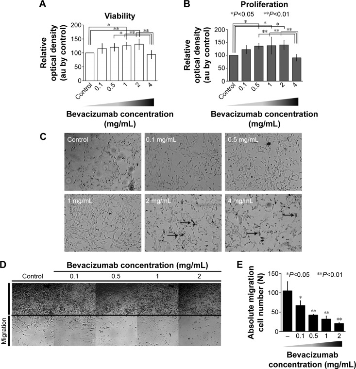
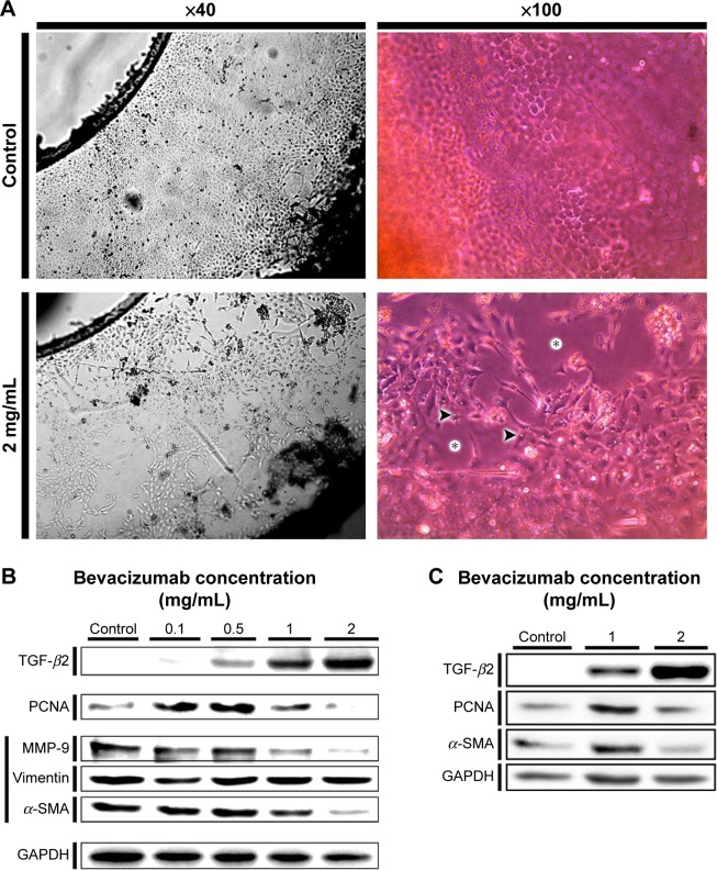
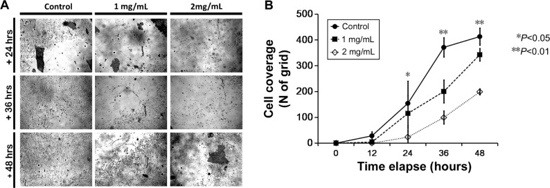
The molecular and cellular effects of anti-vascular endothelial growth factor monoclonal antibody (bevacizumab) on lens epithelial cells (LECs) were examined using both an immortalized human lens epithelial cell line and a porcine capsular bag model. After treatment with various concentrations of bevacizumab, cell viability and proliferation patterns were evaluated using the water-soluble tetrazolium salt assay and 5-bromo-2'-deoxyuridine enzyme-linked immunosorbent assay, respectively. The scratch assay and Western blot analysis were employed to validate the cell migration pattern and altered expression levels of signaling molecules related to the epithelial-mesenchymal transition (EMT). Application of bevacizumab induced a range of altered cellular events in a concentration-dependent manner. A 0.1-2 mg/mL concentration demonstrated dose-dependent increase in proliferation and viability of LECs. However, 4 mg/mL decreased cell proliferation and viability. Cell migrations displayed dose-dependent retardation from 0.1 mg/mL bevacizumab treatment. Transforming growth factor-β2 expression was markedly increased in a dose-dependent manner, and α-smooth muscle actin, matrix metalloproteinase-9, and vimentin expression levels showed dose-dependent changes in a B3 cell line. Microscopic observation of porcine capsular bag revealed changes in cellular morphology and a decline in cell density compared to the control after 2 mg/mL treatment. The central aspect of posterior capsule showed delayed confluence, and the factors related to EMT revealed similar expression patterns to those identified in the cell line. Based on these results, bevacizumab modulates the proliferation and viability of LECs and induces morphological alterations through the modulation of expression patterns of specific factors related to the EMT.

摘要

使用永生化人晶状体上皮细胞系和猪囊袋模型,研究了抗血管内皮生长因子单克隆抗体(贝伐单抗)对晶状体上皮细胞(LECs)的分子和细胞效应。用不同浓度的贝伐单抗处理后,分别使用水溶性四氮唑盐法和5-溴-2'-脱氧尿苷酶联免疫吸附测定法评估细胞活力和增殖模式。采用划痕试验和蛋白质印迹分析来验证细胞迁移模式以及与上皮-间质转化(EMT)相关的信号分子表达水平的变化。贝伐单抗的应用以浓度依赖性方式诱导了一系列细胞事件的改变。0.1-2mg/mL的浓度显示LECs的增殖和活力呈剂量依赖性增加。然而,4mg/mL降低了细胞增殖和活力。从0.1mg/mL贝伐单抗处理开始,细胞迁移显示出剂量依赖性延迟。在B3细胞系中,转化生长因子-β2的表达以剂量依赖性方式显著增加,α-平滑肌肌动蛋白、基质金属蛋白酶-9和波形蛋白的表达水平显示出剂量依赖性变化。猪囊袋的显微镜观察显示,与2mg/mL处理后的对照相比,细胞形态发生了变化,细胞密度下降。后囊中央部分显示融合延迟,与EMT相关的因子显示出与细胞系中鉴定的相似表达模式。基于这些结果,贝伐单抗通过调节与EMT相关的特定因子的表达模式来调节LECs的增殖和活力,并诱导形态学改变。

https://cdn.ncbi.nlm.nih.gov/pmc/blobs/02c9/4935105/a16eaf2bdde2/opth-10-1167Fig3.jpg
https://cdn.ncbi.nlm.nih.gov/pmc/blobs/02c9/4935105/6ae9a10bdb32/opth-10-1167Fig1.jpg
https://cdn.ncbi.nlm.nih.gov/pmc/blobs/02c9/4935105/e5fae9f13fbf/opth-10-1167Fig2.jpg
https://cdn.ncbi.nlm.nih.gov/pmc/blobs/02c9/4935105/a16eaf2bdde2/opth-10-1167Fig3.jpg
https://cdn.ncbi.nlm.nih.gov/pmc/blobs/02c9/4935105/6ae9a10bdb32/opth-10-1167Fig1.jpg
https://cdn.ncbi.nlm.nih.gov/pmc/blobs/02c9/4935105/e5fae9f13fbf/opth-10-1167Fig2.jpg
https://cdn.ncbi.nlm.nih.gov/pmc/blobs/02c9/4935105/a16eaf2bdde2/opth-10-1167Fig3.jpg

相似文献

1
Effects of anti-vascular endothelial growth factor monoclonal antibody (bevacizumab) on lens epithelial cells.抗血管内皮生长因子单克隆抗体(贝伐单抗)对晶状体上皮细胞的作用。
Clin Ophthalmol. 2016 Jun 27;10:1167-74. doi: 10.2147/OPTH.S103443. eCollection 2016.
2
LncRNA HOTAIR mediates TGF-β2-induced cell growth and epithelial-mesenchymal transition in human lens epithelial cells.长链非编码 RNA HOTAIR 介导 TGF-β2 诱导的人晶状体上皮细胞的细胞生长和上皮-间充质转化。
Acta Biochim Biophys Sin (Shanghai). 2018 Oct 1;50(10):1028-1037. doi: 10.1093/abbs/gmy101.
3
Bit1-a potential positive regulator of epithelial-mesenchymal transition in lens epithelial cells.Bit1——晶状体上皮细胞上皮-间质转化的潜在正向调节因子。
Graefes Arch Clin Exp Ophthalmol. 2016 Jul;254(7):1311-8. doi: 10.1007/s00417-016-3357-3. Epub 2016 Apr 28.
4
CtBP2 Regulates TGFβ2-Induced Epithelial-Mesenchymal Transition Through Notch Signaling Pathway in Lens Epithelial Cells.CtBP2通过Notch信号通路调控转化生长因子β2诱导的晶状体上皮细胞上皮-间质转化
Curr Eye Res. 2016 Aug;41(8):1057-1063. doi: 10.3109/02713683.2015.1092554. Epub 2015 Dec 17.
5
The effect of anti-TGF-β2 antibody functionalized intraocular lens on lens epithelial cell migration and epithelial-mesenchymal transition.抗 TGF-β2 抗体功能化人工晶状体对晶状体上皮细胞迁移和上皮-间充质转化的影响。
Colloids Surf B Biointerfaces. 2014 Jan 1;113:33-42. doi: 10.1016/j.colsurfb.2013.08.024. Epub 2013 Sep 1.
6
Enhanced EGF receptor-signaling potentiates TGFβ-induced lens epithelial-mesenchymal transition.增强的表皮生长因子受体信号增强 TGFβ 诱导的晶状体上皮间质转化。
Exp Eye Res. 2019 Aug;185:107693. doi: 10.1016/j.exer.2019.107693. Epub 2019 Jun 12.
7
Implication of Smad2 and Smad3 in transforming growth factor-β-induced posterior capsular opacification of human lens epithelial cells.Smad2和Smad3在转化生长因子-β诱导的人晶状体上皮细胞后囊膜混浊中的作用
Curr Eye Res. 2015 Apr;40(4):386-97. doi: 10.3109/02713683.2014.925932. Epub 2014 Jun 9.
8
Resveratrol Inhibits Wound Healing and Lens Fibrosis: A Putative Candidate for Posterior Capsule Opacification Prevention.白藜芦醇抑制伤口愈合和晶状体纤维化:预防后囊混浊的候选药物。
Invest Ophthalmol Vis Sci. 2019 Sep 3;60(12):3863-3877. doi: 10.1167/iovs.18-26248.
9
An in vitro model of posterior capsular opacity: SPARC and TGF-beta2 minimize epithelial-to-mesenchymal transition in lens epithelium.后囊膜混浊的体外模型:富含半胱氨酸的酸性分泌蛋白(SPARC)和转化生长因子-β2(TGF-β2)可使晶状体上皮细胞的上皮-间充质转化降至最低。
Invest Ophthalmol Vis Sci. 2007 Oct;48(10):4679-87. doi: 10.1167/iovs.07-0091.
10
Suppression of human lens epithelial cell proliferation by proteasome inhibition, a potential defense against posterior capsular opacification.蛋白酶体抑制对人晶状体上皮细胞增殖的抑制作用:一种预防后囊膜混浊的潜在防御机制
Invest Ophthalmol Vis Sci. 2006 Oct;47(10):4482-9. doi: 10.1167/iovs.06-0139.

引用本文的文献

1
Incidence of Posterior Capsule Rupture During Phacoemulsification Cataract Surgery Among Patients Treated With Intravitreal Bevacizumab Injections.玻璃体腔注射贝伐单抗治疗的患者在白内障超声乳化手术中后囊破裂的发生率。
Cureus. 2024 Sep 26;16(9):e70297. doi: 10.7759/cureus.70297. eCollection 2024 Sep.
2
[Lens injury as a complication of intravitreal medication injection].[玻璃体腔内药物注射并发症之晶状体损伤]
Ophthalmologie. 2024 May;121(5):385-390. doi: 10.1007/s00347-024-01997-8. Epub 2024 Feb 16.
3
Therapeutic candidates for keloid scars identified by qualitative review of scratch assay research for wound healing.

本文引用的文献

1
Experimental lens capsular bag model for posterior capsule opacification.用于后囊膜混浊的实验性晶状体囊袋模型
Cell Tissue Res. 2014 Jul;357(1):101-8. doi: 10.1007/s00441-014-1870-4. Epub 2014 May 6.
2
Efficacy of intralesional bevacizumab administration in primary pterygium: a quantitative analysis.经皮腔内注射贝伐单抗治疗原发性翼状胬肉的疗效:定量分析。
Eye Contact Lens. 2014 Jan;40(1):46-50. doi: 10.1097/ICL.0000000000000004.
3
Permeability of the anterior lens capsule for large molecules and small drugs.前晶状体囊对大分子和小分子药物的通透性。
通过定性评价划痕试验研究鉴定瘢痕疙瘩的治疗候选物,以促进伤口愈合。
PLoS One. 2021 Jun 18;16(6):e0253669. doi: 10.1371/journal.pone.0253669. eCollection 2021.
4
Effects of vascular formation during alveolar bone process morphogenesis in mice.小鼠牙槽骨发育过程中血管形成的影响。
Histochem Cell Biol. 2017 Oct;148(4):435-443. doi: 10.1007/s00418-017-1584-2. Epub 2017 Jun 13.
Curr Eye Res. 2013 Oct;38(10):1057-63. doi: 10.3109/02713683.2013.803288. Epub 2013 Jul 25.
4
Randomized, controlled trial of conjunctival autografting combined with subconjunctival bevacizumab for primary pterygium treatment: 1-year follow-up.结膜自体移植联合结膜下注射贝伐单抗治疗原发性翼状胬肉的随机对照试验:1年随访
Clin Exp Ophthalmol. 2014 Apr;42(3):235-41. doi: 10.1111/ceo.12140. Epub 2013 Jul 5.
5
Ranibizumab is a potential prophylaxis for proliferative vitreoretinopathy, a nonangiogenic blinding disease.雷珠单抗是增殖性玻璃体视网膜病变(一种非血管性致盲性疾病)的潜在预防药物。
Am J Pathol. 2013 May;182(5):1659-70. doi: 10.1016/j.ajpath.2013.01.052. Epub 2013 Apr 9.
6
Altered vascular microenvironment by bevacizumab in diabetic fibrovascular membrane.贝伐珠单抗改变糖尿病性纤维血管膜中的血管微环境。
Retina. 2013 May;33(5):957-63. doi: 10.1097/IAE.0b013e3182753b41.
7
Bevacizumab eye drops delay corneal epithelial wound healing and increase the stromal response to epithelial injury in rats.贝伐珠单抗滴眼液延迟大鼠角膜上皮伤口愈合,并增加基质对上皮损伤的反应。
Clin Exp Ophthalmol. 2013 Sep-Oct;41(7):694-701. doi: 10.1111/ceo.12085. Epub 2013 Apr 11.
8
Lenticular cytoprotection. Part 1: the role of hypoxia inducible factors-1α and -2α and vascular endothelial growth factor in lens epithelial cell survival in hypoxia.晶状体细胞保护。第1部分:缺氧诱导因子-1α和-2α以及血管内皮生长因子在缺氧状态下晶状体上皮细胞存活中的作用。
Mol Vis. 2013;19:1-15. Epub 2013 Jan 2.
9
Effects of subconjunctival ranibizumab in a presensitized rat model of corneal graft.结膜下雷珠单抗对角膜移植致敏大鼠模型的影响。
Exp Eye Res. 2013 Feb;107:74-9. doi: 10.1016/j.exer.2012.11.021. Epub 2012 Dec 7.
10
Bevacizumab modulates epithelial-to-mesenchymal transition in the retinal pigment epithelial cells via connective tissue growth factor up-regulation.贝伐单抗通过上调结缔组织生长因子调节视网膜色素上皮细胞的上皮-间充质转化。
Acta Ophthalmol. 2012 Aug;90(5):e389-98. doi: 10.1111/j.1755-3768.2012.02426.x. Epub 2012 Jun 19.