Wu Jie, Gao Ming, Rice Stephen G, Tsang Candy, Beggs John, Turner Dharshaun, Li Guohui, Yang Bo, Xia Kunkun, Gao Fenfei, Qiu Shenfeng, Liu Qiang, Kerrigan John F
The First Affiliated Hospital of Zhengzhou University, Zhengzhou, Henan 450052, China; Division of Neurology, Barrow Neurological Institute, St. Joseph's Hospital and Medical Center, Phoenix, AZ 85013, USA; Department of Pharmacology, Shantou University of Medical College, Guangdong, Shantou 815041, China.
Division of Neurology, Barrow Neurological Institute, St. Joseph's Hospital and Medical Center, Phoenix, AZ 85013, USA.
EBioMedicine. 2016 Jun;8:96-102. doi: 10.1016/j.ebiom.2016.04.026. Epub 2016 Apr 22.
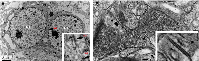
Human hypothalamic hamartoma (HH) is a rare subcortical lesion associated with treatment-resistant epilepsy. Cellular mechanisms responsible for epileptogenesis are unknown. We hypothesized that neuronal gap junctions contribute to epileptogenesis through synchronous activity within the neuron networks in HH tissue. We studied surgically resected HH tissue with Western-blot analysis, immunohistochemistry, electron microscopy, biocytin microinjection of recorded HH neurons, and microelectrode patch clamp recordings with and without pharmacological blockade of gap junctions. Normal human hypothalamus tissue was used as a control. Western blots showed increased expression of both connexin-36 (Cx36) and connexin-43 (Cx43) in HH tissue compared with normal human mammillary body tissue. Immunohistochemistry demonstrated that Cx36 and Cx43 are expressed in HH tissue, but Cx36 was mainly expressed within neuron clusters while Cx43 was mainly expressed outside of neuron clusters. Gap-junction profiles were observed between small HH neurons with electron microscopy. Biocytin injection into single recorded small HH neurons showed labeling of adjacent neurons, which was not observed in the presence of a neuronal gap-junction blocker, mefloquine. Microelectrode field recordings from freshly resected HH slices demonstrated spontaneous ictal/interictal-like discharges in most slices. Bath-application of gap-junction blockers significantly reduced ictal/interictal-like discharges in a concentration-dependent manner, while not affecting the action-potential firing of small gamma-aminobutyric acid (GABA) neurons observed with whole-cell patch-clamp recordings from the same patient's HH tissue. These results suggest that neuronal gap junctions between small GABAergic HH neurons participate in the genesis of epileptic-like discharges. Blockade of gap junctions may be a new therapeutic strategy for controlling seizure activity in HH patients.
人类下丘脑错构瘤(HH)是一种与药物难治性癫痫相关的罕见皮质下病变。导致癫痫发生的细胞机制尚不清楚。我们推测神经元间隙连接通过HH组织中神经元网络内的同步活动促进癫痫发生。我们通过蛋白质免疫印迹分析、免疫组织化学、电子显微镜、对记录的HH神经元进行生物胞素微量注射以及在有无间隙连接药理学阻断的情况下进行微电极膜片钳记录,对手术切除的HH组织进行了研究。正常人类下丘脑组织用作对照。蛋白质免疫印迹显示,与正常人类乳头体组织相比,HH组织中连接蛋白36(Cx36)和连接蛋白43(Cx43)的表达均增加。免疫组织化学表明,Cx36和Cx43在HH组织中表达,但Cx36主要在神经元簇内表达,而Cx43主要在神经元簇外表达。电子显微镜观察到小HH神经元之间存在间隙连接结构。向单个记录的小HH神经元注射生物胞素显示相邻神经元被标记,而在存在神经元间隙连接阻滞剂甲氟喹的情况下未观察到这种情况。对新鲜切除的HH切片进行微电极场记录显示,大多数切片中存在自发性发作期/发作间期样放电。浴用间隙连接阻滞剂以浓度依赖的方式显著减少发作期/发作间期样放电,同时不影响从同一患者的HH组织进行全细胞膜片钳记录时观察到的小γ-氨基丁酸(GABA)神经元的动作电位发放。这些结果表明,小GABA能HH神经元之间的神经元间隙连接参与了癫痫样放电的发生。阻断间隙连接可能是控制HH患者癫痫活动的一种新的治疗策略。