• 文献检索
  • 文档翻译
  • 深度研究
  • 学术资讯
  • Suppr Zotero 插件Zotero 插件
  • 邀请有礼
  • 套餐&价格
  • 历史记录
应用&插件
Suppr Zotero 插件Zotero 插件浏览器插件Mac 客户端Windows 客户端微信小程序
定价
高级版会员购买积分包购买API积分包
服务
文献检索文档翻译深度研究API 文档MCP 服务
关于我们
关于 Suppr公司介绍联系我们用户协议隐私条款
关注我们

Suppr 超能文献

核心技术专利:CN118964589B侵权必究
粤ICP备2023148730 号-1Suppr @ 2026

文献检索

告别复杂PubMed语法,用中文像聊天一样搜索,搜遍4000万医学文献。AI智能推荐,让科研检索更轻松。

立即免费搜索

文件翻译

保留排版,准确专业,支持PDF/Word/PPT等文件格式,支持 12+语言互译。

免费翻译文档

深度研究

AI帮你快速写综述,25分钟生成高质量综述,智能提取关键信息,辅助科研写作。

立即免费体验

使用光学相干断层扫描血管造影术在荧光素血管造影结果正常的普尔夏视网膜病变患者中描绘深层视网膜毛细血管丛中的毛细血管缺失:一例报告

Delineation of capillary dropout in the deep retinal capillary plexus using optical coherence tomography angiography in a patient with Purtscher's retinopathy exhibiting normal fluorescein angiography findings: a case report.

作者信息

Tokimitsu Motoharu, Murata Masako, Toriyama Yuichi, Hirano Takao, Iesato Yasuhiro, Murata Toshinori

机构信息

Department of Ophthalmology, Shinshu University School of Medicine, 3-1-1 Asahi, Matsumoto, Nagano, 390-8621, Japan.

Department of Ophthalmology, Matsumoto Medical Center, National Hospital Organization, Matsumoto, Nagano, Japan.

出版信息

BMC Ophthalmol. 2016 Jul 19;16:113. doi: 10.1186/s12886-016-0298-x.

DOI:10.1186/s12886-016-0298-x
PMID:27430650
原文链接:https://pmc.ncbi.nlm.nih.gov/articles/PMC4950236/
Abstract

BACKGROUND

Fat embolism in the deep retinal capillary plexus is one of the reported mechanisms underlying central/paracentral scotoma in patients with Purtscher's retinopathy. Here we report the clear delineation of capillary dropout in the deep capillary plexus using optical coherence tomography angiography (OCTA) in a chronic case of unexplained scotoma that developed after femoral fracture. The patient exhibited normal fluorescein angiography (FA) findings and a normal retinal appearance.

CASE PRESENTATION

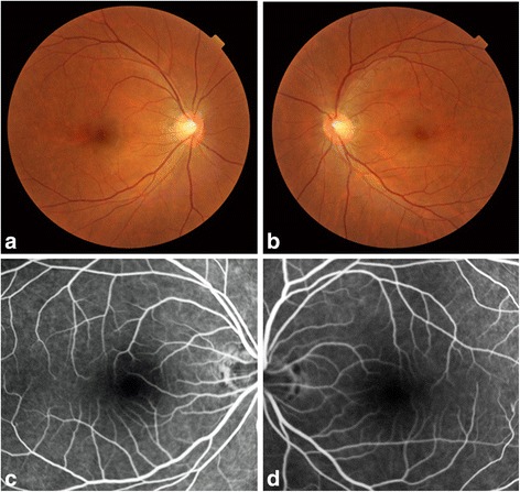
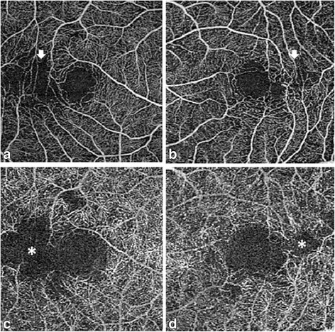
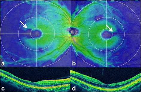
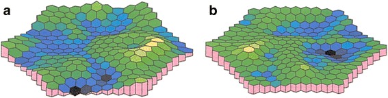
A 42-year-old Japanese man with a history of bilateral, unexplained paracentral scotoma that developed after femoral fracture and pulmonary fat embolism due to a car accident 20 years ago was referred to our outpatient clinic. Initial ophthalmological examination revealed unremarkable retinal findings. Goldmann perimetry, FA, and full field electroretinography showed no pathological changes. Although fat embolism in the retinal vasculature was suspected, psychosomatic visual field defects could not be ruled out. We performed OCTA, which clearly delineated capillary dropout in the deep retinal capillary plexus. A final diagnosis of paracentral acute middle maculopathy secondary to Purtscher's retinopathy was made on the basis of this finding.

CONCLUSIONS

Our findings suggest that OCTA clearly and noninvasively delineates the deep retinal capillary plexus and the superficial capillary plexus. Because conventional FA provides limited depth resolution, capillary dropout restricted within the deep capillary plexus cannot be detected, particularly when the superficial capillary plexus is well preserved. Thus, OCTA can be a useful tool for the detection of capillary dropout in the deep retinal capillary plexus.

摘要

背景

视网膜深层毛细血管丛中的脂肪栓塞是报道的普尔夏视网膜病变患者中心/旁中心暗点的潜在机制之一。在此,我们报告了在一例股骨骨折后出现的不明原因慢性暗点病例中,使用光学相干断层扫描血管造影(OCTA)清晰描绘深层毛细血管丛中毛细血管缺失的情况。该患者荧光素血管造影(FA)结果正常,视网膜外观正常。

病例介绍

一名42岁的日本男性,有双侧不明原因旁中心暗点病史,20年前因车祸导致股骨骨折和肺脂肪栓塞。他被转诊至我们的门诊。初次眼科检查显示视网膜无明显异常。Goldmann视野检查、FA和全视野视网膜电图均未显示病理改变。尽管怀疑视网膜血管存在脂肪栓塞,但不能排除心因性视野缺损。我们进行了OCTA检查,其清晰地描绘了视网膜深层毛细血管丛中的毛细血管缺失。基于这一发现,最终诊断为继发于普尔夏视网膜病变的旁中心急性黄斑病变。

结论

我们的研究结果表明,OCTA能够清晰且无创地描绘视网膜深层毛细血管丛和浅层毛细血管丛。由于传统的FA提供的深度分辨率有限,无法检测到局限于深层毛细血管丛内的毛细血管缺失,尤其是当浅层毛细血管丛保存完好时。因此,OCTA可作为检测视网膜深层毛细血管丛中毛细血管缺失的有用工具。

https://cdn.ncbi.nlm.nih.gov/pmc/blobs/c8b7/4950236/781e6ac0ce83/12886_2016_298_Fig4_HTML.jpg
https://cdn.ncbi.nlm.nih.gov/pmc/blobs/c8b7/4950236/a997bd53714b/12886_2016_298_Fig1_HTML.jpg
https://cdn.ncbi.nlm.nih.gov/pmc/blobs/c8b7/4950236/392ac1a8a985/12886_2016_298_Fig2_HTML.jpg
https://cdn.ncbi.nlm.nih.gov/pmc/blobs/c8b7/4950236/c6a105462136/12886_2016_298_Fig3_HTML.jpg
https://cdn.ncbi.nlm.nih.gov/pmc/blobs/c8b7/4950236/781e6ac0ce83/12886_2016_298_Fig4_HTML.jpg

相似文献

1
Delineation of capillary dropout in the deep retinal capillary plexus using optical coherence tomography angiography in a patient with Purtscher's retinopathy exhibiting normal fluorescein angiography findings: a case report.使用光学相干断层扫描血管造影术在荧光素血管造影结果正常的普尔夏视网膜病变患者中描绘深层视网膜毛细血管丛中的毛细血管缺失:一例报告
BMC Ophthalmol. 2016 Jul 19;16:113. doi: 10.1186/s12886-016-0298-x.
2
Assessing Deep Retinal Capillary Ischemia in Paracentral Acute Middle Maculopathy by Optical Coherence Tomography Angiography.光学相干断层扫描血管造影评估旁中心急性中黄斑病变中的深层视网膜毛细血管缺血情况。
Am J Ophthalmol. 2016 Feb;162:121-132.e1. doi: 10.1016/j.ajo.2015.10.026. Epub 2015 Nov 10.
3
Purtscher's retinopathy case report: short posterior ciliary arteries contribution to radial peripapillary capillary system observed with optical coherence tomography angiography.普尔夏视网膜病变病例报告:光学相干断层扫描血管造影观察到睫状后短动脉对乳头周围放射状毛细血管系统的贡献。
Int Ophthalmol. 2019 Nov;39(11):2661-2665. doi: 10.1007/s10792-019-01086-9. Epub 2019 Mar 9.
4
En Face Optical Coherence Tomography Imaging of Deep Capillary Plexus Abnormalities in Paracentral Acute Middle Maculopathy.中心旁急性黄斑病变中深层毛细血管丛异常的正面光学相干断层扫描成像
Ophthalmic Surg Lasers Imaging Retina. 2015 Oct;46(9):972-5. doi: 10.3928/23258160-20151008-12.
5
OPTICAL COHERENCE TOMOGRAPHY ANGIOGRAPHY SHOWS DEEP CAPILLARY PLEXUS HYPOPERFUSION IN INCOMPLETE CENTRAL RETINAL ARTERY OCCLUSION.光学相干断层扫描血管造影显示不完全性视网膜中央动脉阻塞时深层毛细血管丛灌注不足。
Retin Cases Brief Rep. 2015 Fall;9(4):333-8. doi: 10.1097/ICB.0000000000000211.
6
SPECTRAL DOMAIN VERSUS SWEPT SOURCE OPTICAL COHERENCE TOMOGRAPHY ANGIOGRAPHY OF THE RETINAL CAPILLARY PLEXUSES IN SICKLE CELL MACULOPATHY.镰状细胞性黄斑病变中视网膜毛细血管丛的光谱域与扫频源光学相干断层扫描血管造影术
Retin Cases Brief Rep. 2018;12(2):87-92. doi: 10.1097/ICB.0000000000000448.
7
Spectrum of Retinal Vascular Diseases Associated With Paracentral Acute Middle Maculopathy.与中心旁急性黄斑病变相关的视网膜血管疾病谱
Am J Ophthalmol. 2015 Jul;160(1):26-34.e1. doi: 10.1016/j.ajo.2015.04.004. Epub 2015 Apr 4.
8
TEMPORAL THINNING IN SICKLE CELL RETINOPATHY IS ASSOCIATED WITH DIMINISHED PERFUSION ON OCTA AND DENSE SCOTOMA ON MICROPERIMETERY.镰状细胞视网膜病变中的颞侧变薄与光学相干断层扫描血管造影(OCTA)显示的灌注减少以及微视野检查中的致密暗点相关。
Retin Cases Brief Rep. 2019 Fall;13(4):308-313. doi: 10.1097/ICB.0000000000000610.
9
Assessment of capillary dropout in the superficial retinal capillary plexus by optical coherence tomography angiography in the early stage of diabetic retinopathy.光学相干断层扫描血管造影术评估糖尿病视网膜病变早期浅表视网膜毛细血管丛中的毛细血管缺失情况。
BMC Ophthalmol. 2018 May 8;18(1):113. doi: 10.1186/s12886-018-0778-2.
10
Focal Nonperfusion of Deep Retinal Capillary Plexus in Eyes With Epiretinal Membranes Revealed by Optical Coherence Tomography Angiography.光学相干断层扫描血管造影显示视网膜前膜眼中深层视网膜毛细血管丛的局灶性无灌注
Ophthalmic Surg Lasers Imaging Retina. 2016 May 1;47(5):404-9. doi: 10.3928/23258160-20160419-02.

引用本文的文献

1
Paracentral Acute Middle Maculopathy (PAMM) in Ocular Vascular Diseases-What We Know and Future Perspectives.眼部血管疾病中的黄斑中心凹旁急性中层病变(PAMM)——我们所了解的及未来展望
Vision (Basel). 2025 Mar 3;9(1):19. doi: 10.3390/vision9010019.
2
Electric shock induced Purtscher-like retinopathy.电击相关性酷似普勒歇尔氏视网膜病变。
Indian J Ophthalmol. 2019 Sep;67(9):1497-1500. doi: 10.4103/ijo.IJO_1737_18.
3
Multimodal imaging including optical coherence tomography angiography in patients with type B Niemann-Pick disease.B型尼曼-匹克病患者的多模态成像,包括光学相干断层扫描血管造影术。

本文引用的文献

1
Optical Coherence Tomography Angiography of Deep Capillary Ischemia.深层毛细血管缺血的光学相干断层扫描血管造影术
Dev Ophthalmol. 2016;56:139-45. doi: 10.1159/000442806. Epub 2016 Mar 15.
2
Assessing Deep Retinal Capillary Ischemia in Paracentral Acute Middle Maculopathy by Optical Coherence Tomography Angiography.光学相干断层扫描血管造影评估旁中心急性中黄斑病变中的深层视网膜毛细血管缺血情况。
Am J Ophthalmol. 2016 Feb;162:121-132.e1. doi: 10.1016/j.ajo.2015.10.026. Epub 2015 Nov 10.
3
Spectrum of Retinal Vascular Diseases Associated With Paracentral Acute Middle Maculopathy.
Int Ophthalmol. 2019 Nov;39(11):2545-2552. doi: 10.1007/s10792-019-01102-y. Epub 2019 Apr 11.
4
Multimodal Imaging in Purtscher Retinopathy.普尔夏视网膜病变的多模态成像
Retina. 2018 Jul;38(7):e59-e60. doi: 10.1097/IAE.0000000000002218.
5
Assessment of capillary dropout in the superficial retinal capillary plexus by optical coherence tomography angiography in the early stage of diabetic retinopathy.光学相干断层扫描血管造影术评估糖尿病视网膜病变早期浅表视网膜毛细血管丛中的毛细血管缺失情况。
BMC Ophthalmol. 2018 May 8;18(1):113. doi: 10.1186/s12886-018-0778-2.
6
Purtscher-like retinopathy associated with squamous cell carcinoma of the cervix.与宫颈癌相关的类Purtscher样视网膜病变。
Int Ophthalmol. 2018 Feb;38(1):389-393. doi: 10.1007/s10792-017-0461-2. Epub 2017 Feb 6.
与中心旁急性黄斑病变相关的视网膜血管疾病谱
Am J Ophthalmol. 2015 Jul;160(1):26-34.e1. doi: 10.1016/j.ajo.2015.04.004. Epub 2015 Apr 4.
4
Purtscher-Like Retinopathy: Optical Coherence Tomography and Visual Field Findings.类Purtscher视网膜病变:光学相干断层扫描与视野检查结果
Ophthalmic Surg Lasers Imaging. 2010 Mar 9:1-4. doi: 10.3928/15428877-20100215-51.
5
Sequential multifocal electroretinogram findings in a case of Purtscher-like retinopathy.一例类Purtscher视网膜病变的连续多焦视网膜电图检查结果
Am J Ophthalmol. 2002 Jul;134(1):125-8. doi: 10.1016/s0002-9394(02)01474-5.
6
Unilateral Purtscher's retinopathy.
Ophthalmology. 1980 Nov;87(11):1096-1105. doi: 10.1016/s0161-6420(80)35113-0.
7
Complement-induced granulocyte aggregation: an unsuspected mechanism of disease.补体诱导的粒细胞聚集:一种未被察觉的疾病机制。
N Engl J Med. 1980 Apr 3;302(14):789-94. doi: 10.1056/NEJM198004033021407.
8
Post-traumatic fat embolism located at the retina.创伤后脂肪栓塞位于视网膜。
Acta Ophthalmol (Copenh). 1968;46(2):218-22. doi: 10.1111/j.1755-3768.1968.tb05181.x.
9
Monocular Purtscher's retinopathy. A fluorescein angiographic study.单眼普尔夏视网膜病变。一项荧光素血管造影研究。
Arch Ophthalmol. 1971 Apr;85(4):480-4. doi: 10.1001/archopht.1971.00990050482016.