Sawada Kosaku, Fujioka-Kobayashi Masako, Kobayashi Eizaburo, Brömme Jens O, Schaller Benoit, Miron Richard J
Department of Cranio Maxillofacial Surgery, Inselspital, University of Bern, Bern, Switzerland.
The Nippon Dental University, School of Life Dentistry at Niigata, Advanced Research Center, Niigata, Japan.
BMC Oral Health. 2016 Jul 4;17(1):4. doi: 10.1186/s12903-016-0241-9.
High dose radiation therapy is commonly used in maxillofacial surgeries to treat a number of head and neck tumors. Despite its widespread use, little information is available regarding the effects of irradiation on bone cell viability and release of growth factors following dose-dependent irradiation.
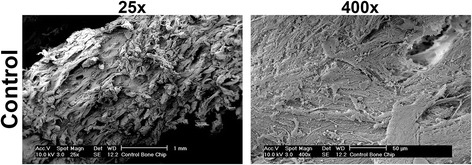
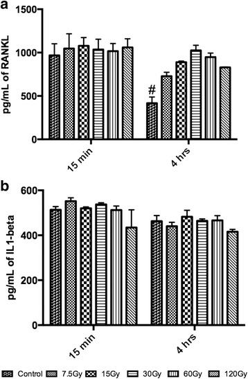
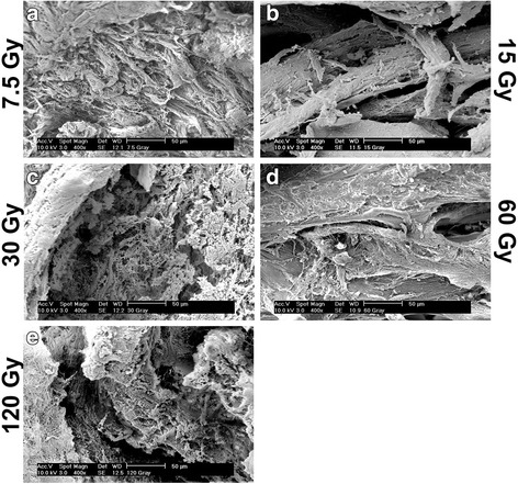
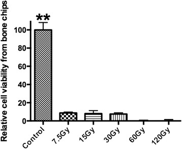
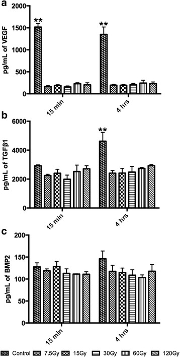
Bone samples were collected from porcine mandibular cortical bone and irradiated at doses of 0, 7.5, 15, 30, 60 and 120 Grays. Thereafter, cell viability was quantified, and the release of growth factors including TGFβ1, BMP2, VEGF, IL1β and RANKL were investigated over time.
It was observed that at only 7.5Gy of irradiation, over 85 % of cells were non-vital and by 60 Gy, all cells underwent apoptosis. Furthermore, over a 7-fold decrease in VEGF and a 2-fold decrease in TGFβ1 were observed following irradiation at all tested doses. Little change was observed for BMP2 and IL1β whereas RANKL was significantly increased for all irradiated samples.
These results demonstrate the pronounced effects of irradiation on bone-cell vitality and subsequent release of growth factors. Interestingly, the largest observed change in gene expression was the 7-fold decrease in VEGF protein following irradiation. Future research aimed at improving our understanding of bone following irradiation is necessary to further improve future clinical treatments.
高剂量放射治疗常用于颌面外科手术,以治疗多种头颈部肿瘤。尽管其应用广泛,但关于剂量依赖性照射后辐射对骨细胞活力和生长因子释放的影响,目前所知甚少。
从猪下颌骨皮质骨采集骨样本,分别用0、7.5、15、30、60和120格雷的剂量进行照射。此后,对细胞活力进行定量分析,并随时间研究包括转化生长因子β1(TGFβ1)、骨形态发生蛋白2(BMP2)、血管内皮生长因子(VEGF)、白细胞介素1β(IL1β)和核因子κB受体活化因子配体(RANKL)在内的生长因子的释放情况。
观察到仅在7.5格雷照射时,超过85%的细胞失去活力,而在60格雷照射时,所有细胞均发生凋亡。此外,在所有测试剂量照射后,VEGF下降超过7倍,TGFβ1下降2倍。BMP2和IL1β变化不大,而所有照射样本的RANKL均显著增加。
这些结果证明了辐射对骨细胞活力以及随后生长因子释放的显著影响。有趣的是,观察到的基因表达最大变化是照射后VEGF蛋白下降7倍。有必要开展进一步研究,以增进我们对辐射后骨骼情况的了解,从而进一步改善未来的临床治疗。