Byrne Eamon F X, Sircar Ria, Miller Paul S, Hedger George, Luchetti Giovanni, Nachtergaele Sigrid, Tully Mark D, Mydock-McGrane Laurel, Covey Douglas F, Rambo Robert P, Sansom Mark S P, Newstead Simon, Rohatgi Rajat, Siebold Christian
Division of Structural Biology, Wellcome Trust Centre for Human Genetics, University of Oxford, Oxford, UK.
Departments of Biochemistry and Medicine, Stanford University School of Medicine, Stanford, California, United States of America.
Nature. 2016 Jul 28;535(7613):517-522. doi: 10.1038/nature18934. Epub 2016 Jul 20.
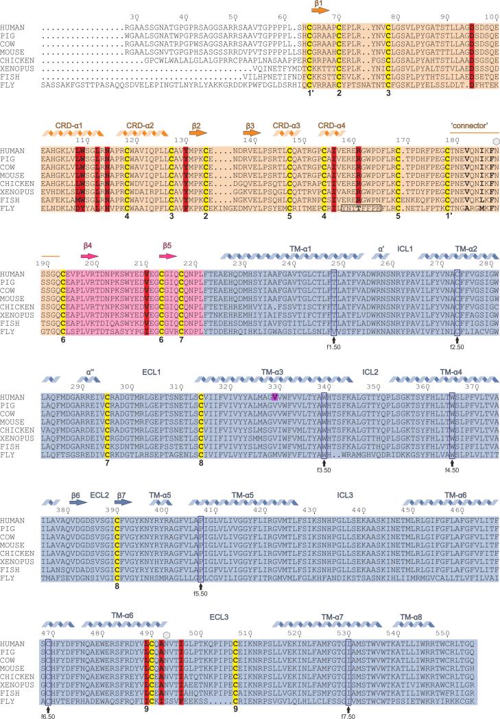
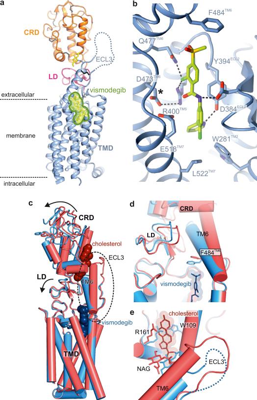
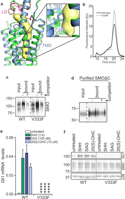
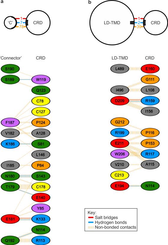
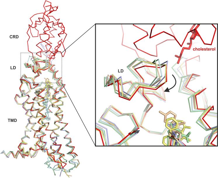
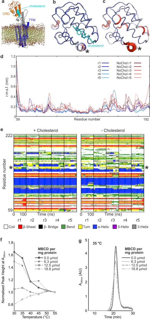
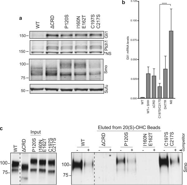
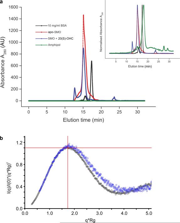
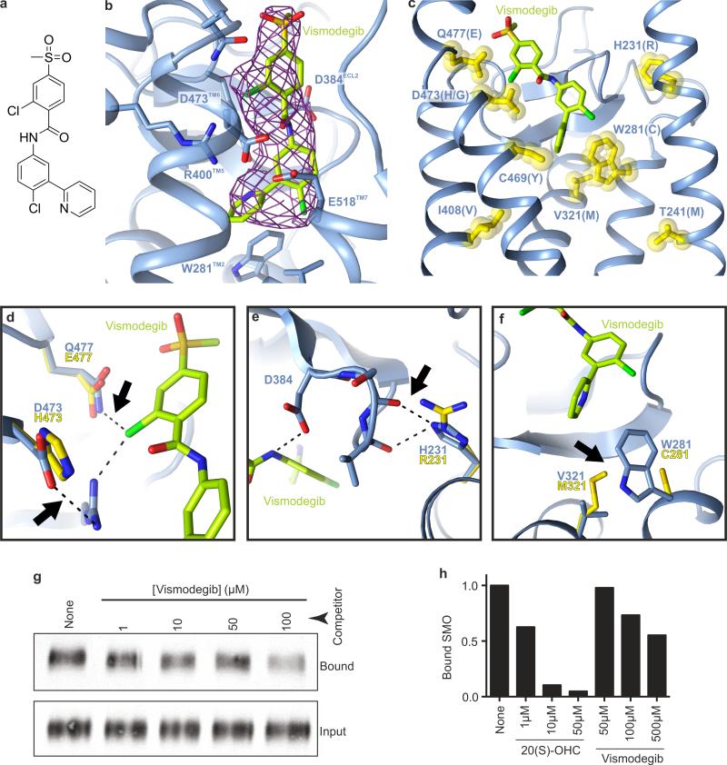
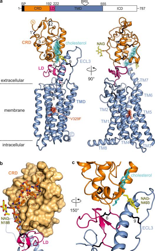
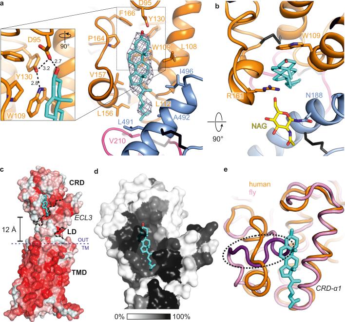
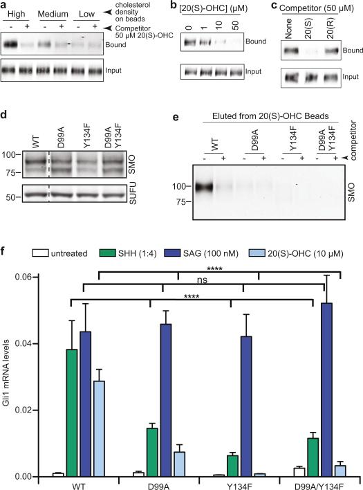
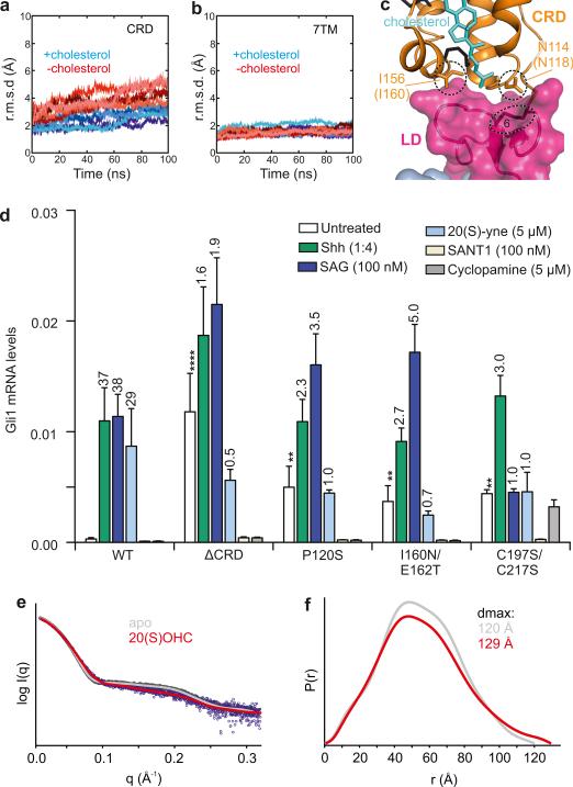
Developmental signals of the Hedgehog (Hh) and Wnt families are transduced across the membrane by Frizzledclass G-protein-coupled receptors (GPCRs) composed of both a heptahelical transmembrane domain (TMD) and an extracellular cysteine-rich domain (CRD). How the large extracellular domains of GPCRs regulate signalling by the TMD is unknown. We present crystal structures of the Hh signal transducer and oncoprotein Smoothened, a GPCR that contains two distinct ligand-binding sites: one in its TMD and one in the CRD. The CRD is stacked a top the TMD, separated by an intervening wedge-like linker domain. Structure-guided mutations show that the interface between the CRD, linker domain and TMD stabilizes the inactive state of Smoothened. Unexpectedly, we find a cholesterol molecule bound to Smoothened in the CRD binding site. Mutations predicted to prevent cholesterol binding impair the ability of Smoothened to transmit native Hh signals. Binding of a clinically used antagonist, vismodegib, to the TMD induces a conformational change that is propagated to the CRD, resulting in loss of cholesterol from the CRD-linker domain-TMD interface. Our results clarify the structural mechanism by which the activity of a GPCR is controlled by ligand-regulated interactions between its extracellular and transmembrane domains.
刺猬索尼克(Hh)和Wnt家族的发育信号通过由七螺旋跨膜结构域(TMD)和富含半胱氨酸的细胞外结构域(CRD)组成的卷曲蛋白类G蛋白偶联受体(GPCR)跨膜传导。GPCR的大细胞外结构域如何调节TMD的信号传导尚不清楚。我们展示了Hh信号转导蛋白和癌蛋白 smoothened(一种GPCR)的晶体结构,它包含两个不同的配体结合位点:一个在其TMD中,另一个在CRD中。CRD堆叠在TMD上方,由一个中间的楔形连接结构域隔开。结构导向突变表明,CRD、连接结构域和TMD之间的界面稳定了smoothened的非活性状态。出乎意料的是,我们在CRD结合位点发现一个胆固醇分子与smoothened结合。预测可阻止胆固醇结合的突变会损害smoothened传递天然Hh信号的能力。临床使用的拮抗剂维莫德吉与TMD结合会诱导构象变化,并传播至CRD,导致胆固醇从CRD-连接结构域-TMD界面丢失。我们的结果阐明了一种GPCR的活性通过其细胞外和跨膜结构域之间的配体调节相互作用来控制的结构机制。