Koirala Pratistha, Roth Michael E, Gill Jonathan, Piperdi Sajida, Chinai Jordan M, Geller David S, Hoang Bang H, Park Amy, Fremed Michael A, Zang Xingxing, Gorlick Richard
Department of Molecular Pharmacology, Albert Einstein College of Medicine, Bronx, NY, USA.
Division of Pediatric Hematology, Oncology, Marrow &Blood Cell Transplantation, Children's Hospital at Montefiore, Albert Einstein College of Medicine, Bronx, NY, USA.
Sci Rep. 2016 Jul 26;6:30093. doi: 10.1038/srep30093.
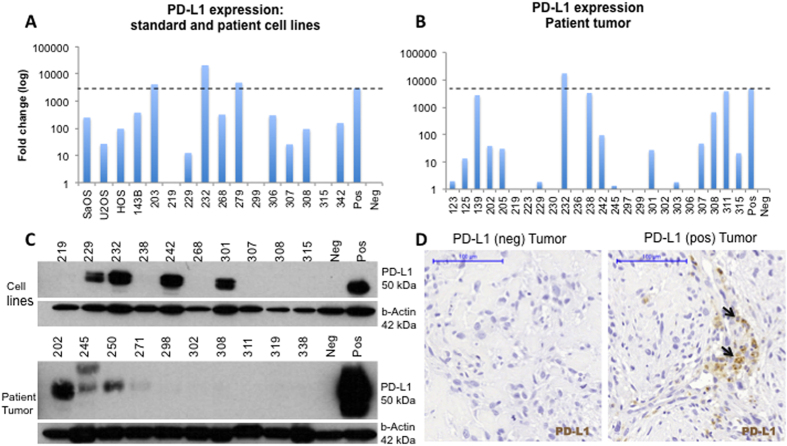
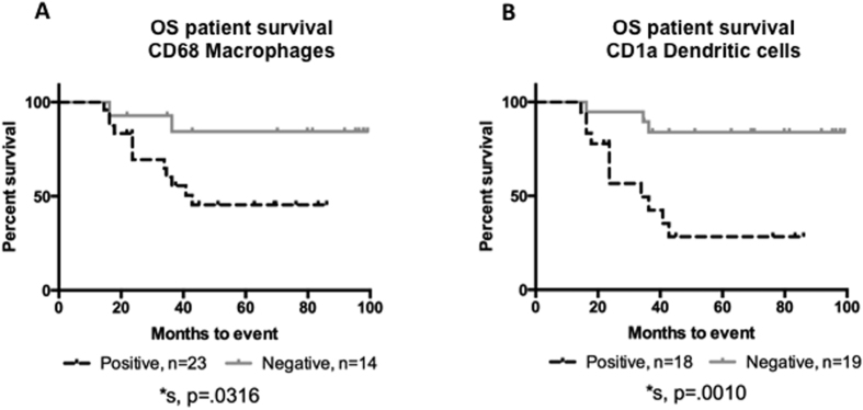
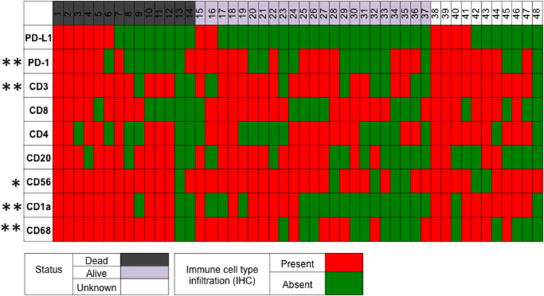
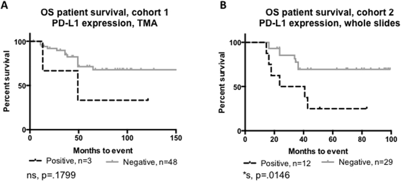
Osteosarcoma patient survival has remained stagnant for 30 years. Novel therapeutic approaches are needed to improve outcomes. We examined the expression of Programmed Death Ligand 1 (PD-L1) and defined the tumor immune microenvironment to assess the prognostic utility in osteosarcoma. PD-L1 expression in osteosarcoma was examined in two patient cohorts using immunohistochemistry (IHC) (n = 48, n = 59) and expression was validated using quantitative real time PCR (n = 21) and western blotting (n = 9). IHC was used to determine the presence of tumor infiltrating lymphocytes and antigen-presenting cells (APCs) in the tumor. Expression of PD-L1 was correlated with immune cell infiltration and event-free-survival (EFS). The 25% of primary osteosarcoma tumors that express PD-L1 were more likely to contain cells that express PD-1 than PD-L1 negative tumors (91.7% vs 47.2%, p = 0.002). Expression of PD-L1 was significantly associated with the presence of T cells, dendritic cells, and natural killer cells. Although all immune cell types examined were present in osteosarcoma samples, only infiltration by dendritic cells (28.3% vs. 83.9%, p = 0.001) and macrophages (45.5% vs. 84.4%, p = 0.031) were associated with worse five-year-EFS. PD-L1 expression was significantly associated with poorer five-year-EFS (25.0%. vs. 69.4%, p = 0.014). Further studies in osteosarcoma are needed to determine if targeting the PD-L1:PD-1 axis improves survival.
骨肉瘤患者的生存率在30年里一直停滞不前。需要新的治疗方法来改善治疗效果。我们检测了程序性死亡配体1(PD-L1)的表达,并定义了肿瘤免疫微环境,以评估其在骨肉瘤中的预后效用。使用免疫组织化学(IHC)在两个患者队列中检测骨肉瘤中PD-L1的表达(n = 48,n = 59),并使用定量实时PCR(n = 21)和蛋白质免疫印迹法(n = 9)验证表达情况。IHC用于确定肿瘤中肿瘤浸润淋巴细胞和抗原呈递细胞(APC)的存在。PD-L1的表达与免疫细胞浸润和无事件生存期(EFS)相关。与PD-L1阴性肿瘤相比,表达PD-L1的25%原发性骨肉瘤肿瘤更有可能含有表达PD-1的细胞(91.7%对47.2%,p = 0.002)。PD-L1的表达与T细胞、树突状细胞和自然杀伤细胞的存在显著相关。尽管在骨肉瘤样本中检测到了所有类型的免疫细胞,但只有树突状细胞浸润(28.3%对83.9%,p = 0.001)和巨噬细胞浸润(45.5%对84.4%,p = 0.031)与较差的五年EFS相关。PD-L1表达与较差的五年EFS显著相关(25.0%对69.4%,p = 0.014)。需要对骨肉瘤进行进一步研究,以确定靶向PD-L1:PD-1轴是否能提高生存率。