Li Liang, Liang Chaozhao
Department of Ultrasound, The First Affiliated Hospital of Anhui Medical University, Hefei, Anhui, China (mainland).
Department of Urology, The First Affiliated Hospital of Anhui Medical University, Hefei, Anhui, China (mainland).
Med Sci Monit. 2016 Jul 26;22:2643-7. doi: 10.12659/msm.898109.
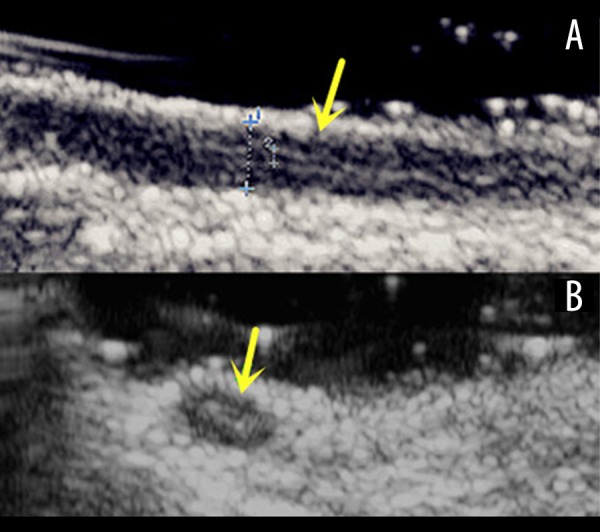
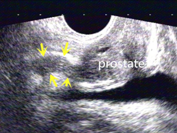
BACKGROUND Congenital absence of the vas deferens is an important cause of obstructive azoospermia, and the lack of an imaging diagnostic test is a critical problem. The aim of this study is to discuss the use of ultrasonography in congenital absence of vas deferens, including dysplasia of the epididymis and the seminal vesical. MATERIAL AND METHODS Five fresh spermatic cord specimens were detected by ultrasonography (US) to evaluate the image of the spermatic cord segment of the vas deferens. Fifty normal males had scrotal US to confirm whether the normal spermatic cord segment of the vas deferens can be detected and to measure the internal and external diameter on the long axis view. Forty-six males clinically diagnosed as having congenital absence of vas deferens underwent scrotal US to evaluate the spermatic cord segment of the vas deferens and the epididymis. The seminal vesicals were detected with transrectal ultrasonography. We evaluated images of the vas deferens, epididymis, and seminal vesical. RESULTS Scrotal ultrasonography can distinguish the vas deferens from the other cord-like structures in the spermatic cord, and the vas deferens has a characteristic image. Scrotal ultrasonography detected all 50 normal males and measured the diameter. No statistically significant difference was found between the left and right measurements. In the 46 patients, the following anomalies were observed: 1) 42 cases of congenital bilateral absence of vas deferens; 2) 2 cases of congenital unilateral absence of the vas deferens; and 3) 1 case of congenital segmental absence of the vas deferens. All 46 cases were accompanied with epididymis and seminal vesical anomalies. CONCLUSIONS The spermatic cord segment of the vas deferens can be detected by US, which is a valuable tool in diagnosis of congenital absence of the vas deferens. Seminal vesical and epididymis anomalies often associated with congenital absence of the vas deferens were revealed by ultrasonography.
背景 先天性输精管缺如是梗阻性无精子症的重要原因,而缺乏影像学诊断检查是一个关键问题。本研究的目的是探讨超声检查在先天性输精管缺如中的应用,包括附睾和精囊发育异常情况。
材料与方法 对5个新鲜精索标本进行超声检查,以评估输精管精索段的图像。对50名正常男性进行阴囊超声检查,以确认是否能检测到正常的输精管精索段,并在长轴视图上测量其内径和外径。对46例临床诊断为先天性输精管缺如的男性进行阴囊超声检查,以评估输精管精索段和附睾情况。经直肠超声检查精囊。我们评估了输精管、附睾和精囊的图像。
结果 阴囊超声检查可将输精管与精索内其他条索状结构区分开来,输精管具有特征性图像。阴囊超声检查检测出了所有50名正常男性并测量了直径。左右测量值之间未发现统计学上的显著差异。在46例患者中,观察到以下异常情况:1)42例先天性双侧输精管缺如;2)2例先天性单侧输精管缺如;3)1例先天性节段性输精管缺如。所有46例均伴有附睾和精囊异常。
结论 超声检查可检测输精管精索段,这是诊断先天性输精管缺如的一种有价值的工具。超声检查揭示了常与先天性输精管缺如相关的精囊和附睾异常情况。