Yu Lin, Liu Jinguo, Lao I Weng, Luo Zhiguo, Wang Jian
Department of Pathology, Fudan University Shanghai Cancer Center, Fudan University, 270 Dong An Road, Shanghai, 200032, China.
Department of Oncology, Shanghai Medical College, Fudan University, Shanghai, 200032, China.
Diagn Pathol. 2016 Jul 27;11(1):67. doi: 10.1186/s13000-016-0517-z.
To explore the clinical characteristics and pathological features of epithelioid inflammatory myofibroblastic sarcoma (EIMS) with emphasis on the diagnostic spectrum.
The clinical data and histological features in 5 additional cases of EIMS were retrospectively reviewed. Immunohistochemical study and interphase fluorescence in situ hybridization (FISH) analysis were carried out.
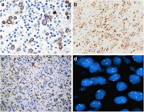
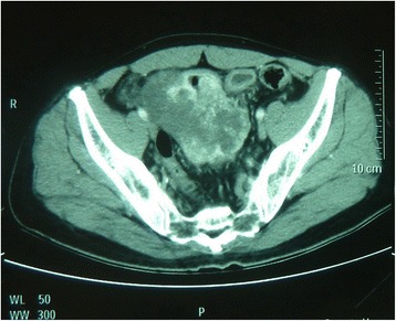
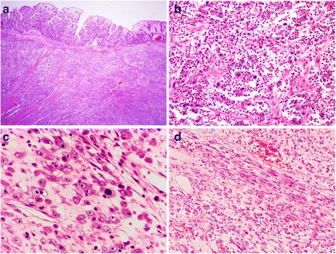
There were 2 males and 3 females with age at presentation ranging from 15 to 58 years (mean, 37 years). All 5 tumors were intra-abdominal with 2 arising in the mesentery and 1 each in the omentum, rectum and transverse colon. The tumor size ranged from 5 to 20 cm in maximum diameter (mean, 10.7 cm). Histologically, all 5 tumors were composed predominantly of large epithelioid cells possessing vesicular nuclei, prominent nucleoli, and amphophilic cytoplasm. Mitotic figures were easily identified (mean, 20/10HPF). Tumor cells were arranged in clusters or sheets embedded in a myxoid stroma containing prominent neutrophils. A minor component of spindle cells was present in focal areas. By immunohistochemistry, all 5 cases were positive for anaplastic lymphoma kinase (ALK) with a nuclear membrane pattern in 4 and cytoplasmic staining with perinuclear accentuation in 1. Besides ALK, tumor cells stained variably for desmin (4/5), alpha smooth muscle actin (2/5), muscle-specific actin (1/2) and pan-cytokeratin (1/4). FISH analysis demonstrated the presence of ALK rearrangement in all 5 cases. Of 5 patients, 3 developed local recurrence, 1 died of disease 8 months after surgery.
EIMS represents a highly aggressive variant of inflammatory myofibroblastic tumor characterized by epithelioid morphology, prominent neutrophilic infiltrate, and nuclear membrane staining of ALK with ALK rearrangement. As patients with ALK-rearrangement tumors may benefit from targeted therapy, accurate diagnosis of EIMS is very important. Familiar with the characteristic features of EIMS will help pathologists avoid misdiagnosing the tumor as other malignancies.
探讨上皮样炎性肌纤维母细胞肉瘤(EIMS)的临床特征和病理特征,重点关注其诊断范围。
回顾性分析另外5例EIMS患者的临床资料和组织学特征。进行免疫组织化学研究和间期荧光原位杂交(FISH)分析。
5例患者中,男性2例,女性3例,发病年龄为15至58岁(平均37岁)。所有5个肿瘤均位于腹腔内,2个起源于肠系膜,大网膜、直肠和横结肠各1个。肿瘤最大直径为5至20厘米(平均10.7厘米)。组织学上,所有5个肿瘤主要由大的上皮样细胞组成,这些细胞具有泡状核、明显的核仁以及嗜双色性细胞质。有丝分裂象容易识别(平均20/10高倍视野)。肿瘤细胞呈簇状或片状排列,包埋于含有大量中性粒细胞的黏液样基质中。局部区域存在少量梭形细胞成分。免疫组织化学检测显示,所有5例间变性淋巴瘤激酶(ALK)均呈阳性,其中4例为核膜型,1例为细胞质染色伴核周增强。除ALK外,肿瘤细胞对结蛋白(4/5)、α平滑肌肌动蛋白(2/5)、肌肉特异性肌动蛋白(1/2)和泛细胞角蛋白(1/4)的染色各不相同。FISH分析显示所有5例均存在ALK重排。5例患者中,3例出现局部复发,1例术后8个月死于疾病。
EIMS是炎性肌纤维母细胞瘤的一种高度侵袭性变体,其特征为上皮样形态、显著的中性粒细胞浸润以及ALK核膜染色伴ALK重排。由于ALK重排肿瘤患者可能从靶向治疗中获益,准确诊断EIMS非常重要。熟悉EIMS的特征将有助于病理学家避免将该肿瘤误诊为其他恶性肿瘤。