• 文献检索
  • 文档翻译
  • 深度研究
  • 学术资讯
  • Suppr Zotero 插件Zotero 插件
  • 邀请有礼
  • 套餐&价格
  • 历史记录
应用&插件
Suppr Zotero 插件Zotero 插件浏览器插件Mac 客户端Windows 客户端微信小程序
定价
高级版会员购买积分包购买API积分包
服务
文献检索文档翻译深度研究API 文档MCP 服务
关于我们
关于 Suppr公司介绍联系我们用户协议隐私条款
关注我们

Suppr 超能文献

核心技术专利:CN118964589B侵权必究
粤ICP备2023148730 号-1Suppr @ 2026

文献检索

告别复杂PubMed语法,用中文像聊天一样搜索,搜遍4000万医学文献。AI智能推荐,让科研检索更轻松。

立即免费搜索

文件翻译

保留排版,准确专业,支持PDF/Word/PPT等文件格式,支持 12+语言互译。

免费翻译文档

深度研究

AI帮你快速写综述,25分钟生成高质量综述,智能提取关键信息,辅助科研写作。

立即免费体验

利用乙醇固定和大面积光子计数探测器对离体小鼠软组织器官进行高对比度X射线显微放射成像和显微CT成像。

High-contrast X-ray micro-radiography and micro-CT of ex-vivo soft tissue murine organs utilizing ethanol fixation and large area photon-counting detector.

作者信息

Dudak Jan, Zemlicka Jan, Karch Jakub, Patzelt Matej, Mrzilkova Jana, Zach Petr, Hermanova Zuzana, Kvacek Jiri, Krejci Frantisek

机构信息

Institute of Experimental and Applied Physics, Czech Technical University in Prague, Horska 3a/22, 128 00 Prague, Czech Republic.

Faculty of Biomedical Engineering, Czech Technical University in Prague, Namesti Sitna 3105, 272 01 Kladno, Czech Republic.

出版信息

Sci Rep. 2016 Jul 27;6:30385. doi: 10.1038/srep30385.

DOI:10.1038/srep30385
PMID:27461900
原文链接:https://pmc.ncbi.nlm.nih.gov/articles/PMC4961961/
Abstract

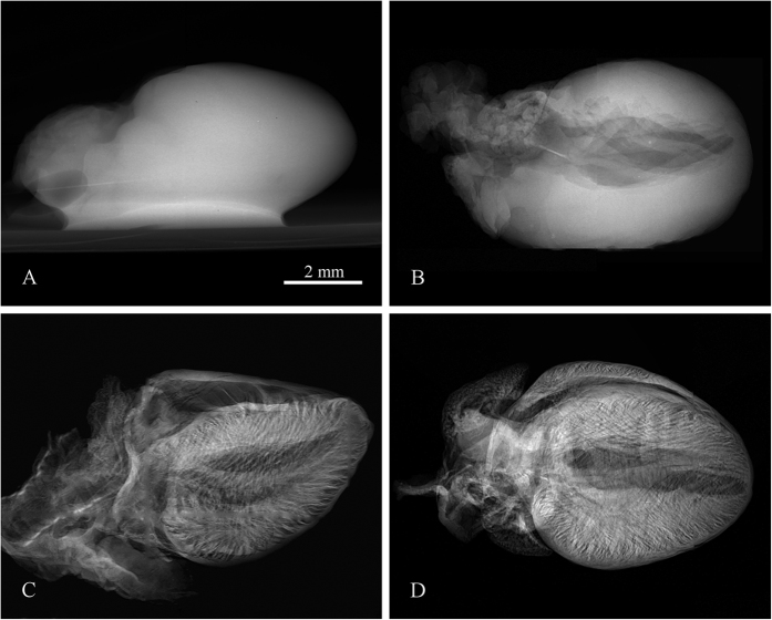
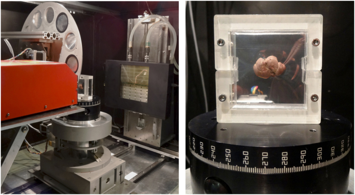
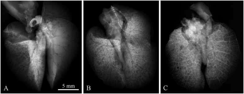
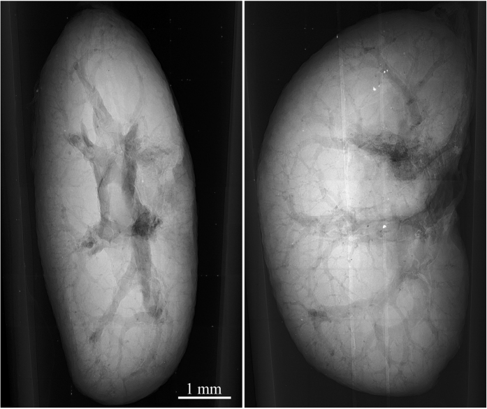
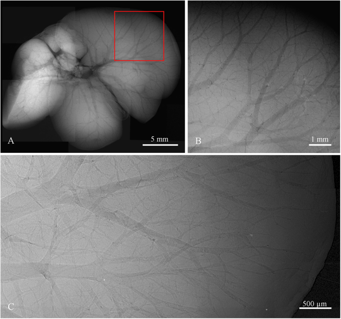
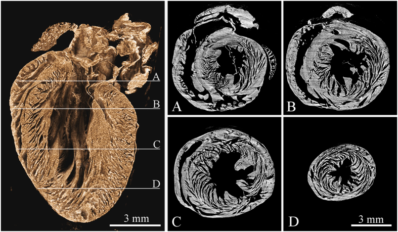
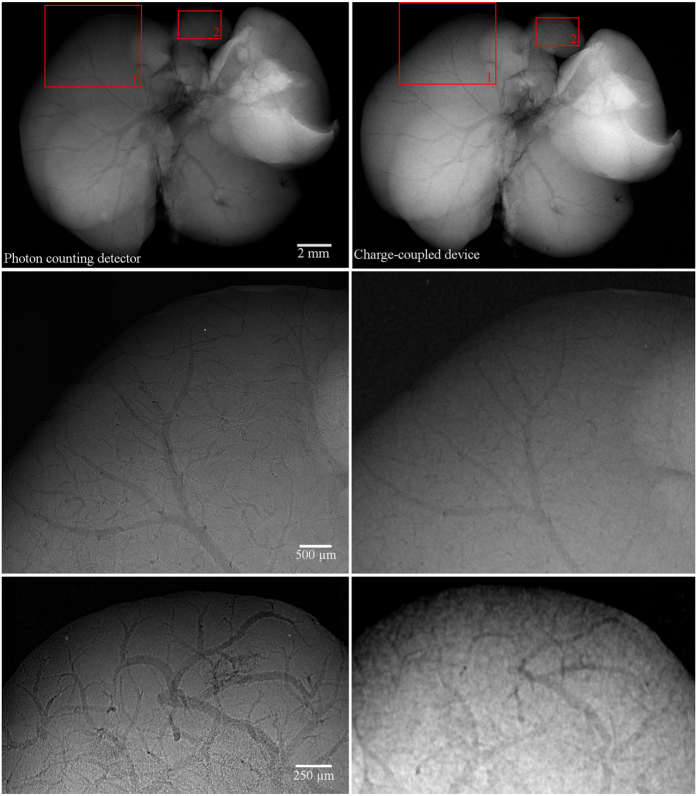
Using dedicated contrast agents high-quality X-ray imaging of soft tissue structures with isotropic micrometre resolution has become feasible. This technique is frequently titled as virtual histology as it allows production of slices of tissue without destroying the sample. The use of contrast agents is, however, often an irreversible time-consuming procedure and despite the non-destructive principle of X-ray imaging, the sample is usually no longer usable for other research methods. In this work we present the application of recently developed large-area photon counting detector for high resolution X-ray micro-radiography and micro-tomography of whole ex-vivo ethanol-preserved mouse organs. The photon counting detectors provide dark-current-free quantum-counting operation enabling acquisition of data with virtually unlimited contrast-to-noise ratio (CNR). Thanks to the very high CNR even ethanol-only preserved soft-tissue samples without addition of any contrast agent can be visualized in great detail. As ethanol preservation is one of the standard steps of tissue fixation for histology, the presented method can open a way for widespread use of micro-CT with all its advantages for routine 3D non-destructive soft-tissue visualisation.

摘要

使用专用造影剂,实现具有各向同性微米分辨率的软组织结构的高质量X射线成像已成为可能。这种技术常被称为虚拟组织学,因为它能够在不破坏样本的情况下生成组织切片。然而,造影剂的使用往往是一个不可逆的耗时过程,并且尽管X射线成像具有无损原理,但样本通常不再适用于其他研究方法。在这项工作中,我们展示了最近开发的大面积光子计数探测器在对整个离体乙醇保存的小鼠器官进行高分辨率X射线显微放射成像和显微断层扫描中的应用。光子计数探测器提供无暗电流的量子计数操作,能够获取具有几乎无限对比度噪声比(CNR)的数据。由于极高的CNR,即使是未添加任何造影剂的仅用乙醇保存的软组织样本也能被非常详细地可视化。由于乙醇保存是组织学中组织固定的标准步骤之一,所提出的方法可以为广泛使用具有其所有优势的微型计算机断层扫描(micro-CT)用于常规三维无损软组织可视化开辟道路。

https://cdn.ncbi.nlm.nih.gov/pmc/blobs/250e/4961961/07120f35aa7f/srep30385-f7.jpg
https://cdn.ncbi.nlm.nih.gov/pmc/blobs/250e/4961961/f0b7d44248c9/srep30385-f1.jpg
https://cdn.ncbi.nlm.nih.gov/pmc/blobs/250e/4961961/c70e685e5f67/srep30385-f2.jpg
https://cdn.ncbi.nlm.nih.gov/pmc/blobs/250e/4961961/78a93bac3bca/srep30385-f3.jpg
https://cdn.ncbi.nlm.nih.gov/pmc/blobs/250e/4961961/3c185895a3e5/srep30385-f4.jpg
https://cdn.ncbi.nlm.nih.gov/pmc/blobs/250e/4961961/6207ee1cefa6/srep30385-f5.jpg
https://cdn.ncbi.nlm.nih.gov/pmc/blobs/250e/4961961/87fa8bb646e8/srep30385-f6.jpg
https://cdn.ncbi.nlm.nih.gov/pmc/blobs/250e/4961961/07120f35aa7f/srep30385-f7.jpg

相似文献

1
High-contrast X-ray micro-radiography and micro-CT of ex-vivo soft tissue murine organs utilizing ethanol fixation and large area photon-counting detector.利用乙醇固定和大面积光子计数探测器对离体小鼠软组织器官进行高对比度X射线显微放射成像和显微CT成像。
Sci Rep. 2016 Jul 27;6:30385. doi: 10.1038/srep30385.
2
Ethanol fixation method for heart and lung imaging in micro-CT.微 CT 中心肺成像的乙醇固定方法。
Jpn J Radiol. 2019 Jun;37(6):500-510. doi: 10.1007/s11604-019-00830-6. Epub 2019 Mar 14.
3
Non-destructive imaging of fragments of historical beeswax seals using high-contrast X-ray micro-radiography and micro-tomography with large area photon-counting detector array.使用带有大面积光子计数探测器阵列的高对比度X射线显微放射成像和显微断层成像技术对历史蜂蜡印章碎片进行无损成像。
Micron. 2016 Dec;91:22-28. doi: 10.1016/j.micron.2016.09.006. Epub 2016 Sep 16.
4
Synchrotron inline phase contrast µCT enables detailed virtual histology of embedded soft-tissue samples with and without staining.同步加速器在线相衬显微CT能够对嵌入的软组织样本进行详细的虚拟组织学分析,无论样本是否经过染色。
J Synchrotron Radiat. 2018 Jul 1;25(Pt 4):1153-1161. doi: 10.1107/S1600577518005489. Epub 2018 May 31.
5
Nanoparticle contrast-enhanced micro-CT: A preclinical tool for the 3D imaging of liver and spleen in longitudinal mouse studies.纳米颗粒对比增强微型计算机断层扫描:一种用于小鼠纵向研究中肝脏和脾脏三维成像的临床前工具。
J Pharmacol Toxicol Methods. 2019 Mar-Apr;96:67-77. doi: 10.1016/j.vascn.2019.02.003. Epub 2019 Feb 7.
6
Synchrotron- and laboratory-based X-ray phase-contrast imaging for imaging mouse articular cartilage in the absence of radiopaque contrast agents.同步辐射和实验室基于 X 射线相衬成像在没有不透射线对比剂的情况下对小鼠关节软骨成像。
Philos Trans A Math Phys Eng Sci. 2014 Jan 27;372(2010):20130127. doi: 10.1098/rsta.2013.0127. Print 2014 Mar 6.
7
Contrast-enhanced photon-counting micro-CT of tumor xenograft models.肿瘤异种移植模型的对比增强光子计数微 CT
Phys Med Biol. 2024 Jul 19;69(15):155011. doi: 10.1088/1361-6560/ad4447.
8
Estimation of mouse organ locations through registration of a statistical mouse atlas with micro-CT images.通过将统计小鼠图谱与 micro-CT 图像进行配准来估计小鼠器官位置。
IEEE Trans Med Imaging. 2012 Jan;31(1):88-102. doi: 10.1109/TMI.2011.2165294. Epub 2011 Aug 18.
9
Dual-energy micro-CT for quantifying the time-course and staining characteristics of ex-vivo animal organs treated with iodine- and gadolinium-based contrast agents.双能 micro-CT 定量分析碘基和钆基对比剂处理的离体动物器官的时程和染色特征。
Sci Rep. 2017 Dec 12;7(1):17387. doi: 10.1038/s41598-017-17064-z.
10
Tilted angle CZT detector for photon counting/energy weighting x-ray and CT imaging.用于光子计数/能量加权X射线和CT成像的倾斜角碲锌镉探测器。
Phys Med Biol. 2006 Sep 7;51(17):4267-87. doi: 10.1088/0031-9155/51/17/010. Epub 2006 Aug 15.

引用本文的文献

1
The use of ethanol as contrast enhancer in synchrotron X-ray phase-contrast imaging leads to heterogeneous myocardial tissue shrinkage: a case report.同步加速器X射线相衬成像中使用乙醇作为对比增强剂导致心肌组织不均匀收缩:一例报告。
J Synchrotron Radiat. 2025 Jan 1;32(Pt 1):200-209. doi: 10.1107/S1600577524010221.
2
Phase contrast micro-CT with adjustable in-slice spatial resolution at constant magnification.具有可调切片内空间分辨率的相衬微 CT,在恒定放大倍数下。
Phys Med Biol. 2024 May 7;69(10):105017. doi: 10.1088/1361-6560/ad4000.
3
Visualization of rat tendon in three dimensions using micro-Computed Tomography.

本文引用的文献

1
High contrast microstructural visualization of natural acellular matrices by means of phase-based x-ray tomography.通过基于相位的X射线断层扫描对天然无细胞基质进行高对比度微观结构可视化。
Sci Rep. 2015 Dec 14;5:18156. doi: 10.1038/srep18156.
2
Three-dimensional non-destructive soft-tissue visualization with X-ray staining micro-tomography.利用X射线染色显微断层扫描技术进行三维无损软组织可视化。
Sci Rep. 2015 Sep 25;5:14088. doi: 10.1038/srep14088.
3
Contrast agents for preclinical targeted X-ray imaging.用于临床前靶向 X 射线成像的造影剂。
使用微型计算机断层扫描对大鼠肌腱进行三维可视化。
MethodsX. 2024 Jan 10;12:102565. doi: 10.1016/j.mex.2024.102565. eCollection 2024 Jun.
4
Optimal Agents for Visualizing Collagen Tissue Microarchitecture Using Contrast-Enhanced MicroCT.使用对比增强显微CT可视化胶原组织微观结构的最佳试剂
Pharmaceuticals (Basel). 2023 Dec 12;16(12):1719. doi: 10.3390/ph16121719.
5
An optimized workflow for microCT imaging of formalin-fixed and paraffin-embedded (FFPE) early equine embryos.优化福尔马林固定石蜡包埋(FFPE)早期马胚胎 microCT 成像的工作流程。
Anat Histol Embryol. 2022 Sep;51(5):611-623. doi: 10.1111/ahe.12834. Epub 2022 Jul 18.
6
Brain virtual histology with X-ray phase-contrast tomography Part I: whole-brain myelin mapping in white-matter injury models.基于X射线相衬断层扫描的脑虚拟组织学 第一部分:白质损伤模型中的全脑髓鞘图谱
Biomed Opt Express. 2022 Feb 23;13(3):1620-1639. doi: 10.1364/BOE.438832. eCollection 2022 Mar 1.
7
A Transparent Nano-Polycrystalline ZnWO Thin-Film Scintillator for High-Resolution X-ray Imaging.用于高分辨率X射线成像的透明纳米多晶ZnWO薄膜闪烁体
ACS Omega. 2021 Nov 22;6(48):33224-33230. doi: 10.1021/acsomega.1c05962. eCollection 2021 Dec 7.
8
A quality optimization approach to image Achilles tendon microstructure by phase-contrast enhanced synchrotron micro-tomography.基于相衬增强同步辐射微层析成像的图像跟腱微结构的质量优化方法。
Sci Rep. 2021 Aug 27;11(1):17313. doi: 10.1038/s41598-021-96589-w.
9
X-ray microtomosynthesis of unstained pathology tissue samples.未染色病理组织样本的 X 射线显微断层摄影术。
J Microsc. 2021 Jul;283(1):9-20. doi: 10.1111/jmi.13003. Epub 2021 Feb 18.
10
Image quality assessment of a photon counting detector in x-ray projection imaging.X射线投影成像中光子计数探测器的图像质量评估
Nucl Instrum Methods Phys Res A. 2019 Sep 21;939:83-88. doi: 10.1016/j.nima.2019.05.054. Epub 2019 May 23.
Adv Drug Deliv Rev. 2014 Sep 30;76:116-133. doi: 10.1016/j.addr.2014.07.013. Epub 2014 Jul 30.
4
Enhanced renal image contrast by ethanol fixation in phase-contrast X-ray computed tomography.通过乙醇固定在相衬X射线计算机断层扫描中增强肾脏图像对比度。
J Synchrotron Radiat. 2014 Jul;21(Pt 4):795-800. doi: 10.1107/S1600577514010558. Epub 2014 Jun 12.
5
In-vivo dark-field and phase-contrast x-ray imaging.体内暗场与相衬X射线成像。
Sci Rep. 2013 Nov 13;3:3209. doi: 10.1038/srep03209.
6
Multimodal imaging of human cerebellum - merging X-ray phase microtomography, magnetic resonance microscopy and histology.多模态人类小脑成像 - 融合 X 射线相衬显微断层成像、磁共振显微镜和组织学。
Sci Rep. 2012;2:826. doi: 10.1038/srep00826. Epub 2012 Nov 9.
7
Emphysema diagnosis using X-ray dark-field imaging at a laser-driven compact synchrotron light source.使用激光驱动紧凑型同步辐射光源的 X 射线暗场成象进行肺气肿诊断。
Proc Natl Acad Sci U S A. 2012 Oct 30;109(44):17880-5. doi: 10.1073/pnas.1206684109. Epub 2012 Oct 16.
8
Coronary artery wall imaging in mice using osmium tetroxide and micro-computed tomography (micro-CT).使用锇四氧化物和微计算机断层扫描(micro-CT)对小鼠的冠状动脉壁成像。
J Anat. 2012 May;220(5):514-24. doi: 10.1111/j.1469-7580.2012.01483.x. Epub 2012 Feb 23.
9
X-ray microtomography in biology.X 射线显微断层成像技术在生物学中的应用。
Micron. 2012 Feb;43(2-3):104-15. doi: 10.1016/j.micron.2011.10.002. Epub 2011 Oct 10.
10
Imaging heart development using high-resolution episcopic microscopy.使用高分辨率的外照显微镜成像心脏发育。
Curr Opin Genet Dev. 2011 Oct;21(5):573-8. doi: 10.1016/j.gde.2011.07.004. Epub 2011 Sep 4.