Hines Catherine D G, Wang Shubing, Meng Xiangjun, Skinner Julie M, Heinrichs Jon H, Smith Jeffrey G, Boddicker Melissa A
Department of Translational Imaging Biomarkers, MRL (West Point, PA), Merck & Co., Inc., Kenilworth, New Jersey, United States of America.
Department of Biometrics Research, MRL (Rahway, NJ), Merck & Co., Inc., Kenilworth, New Jersey, United States of America.
PLoS One. 2016 Jul 28;11(7):e0160055. doi: 10.1371/journal.pone.0160055. eCollection 2016.
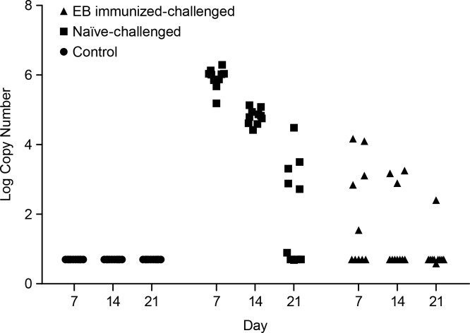
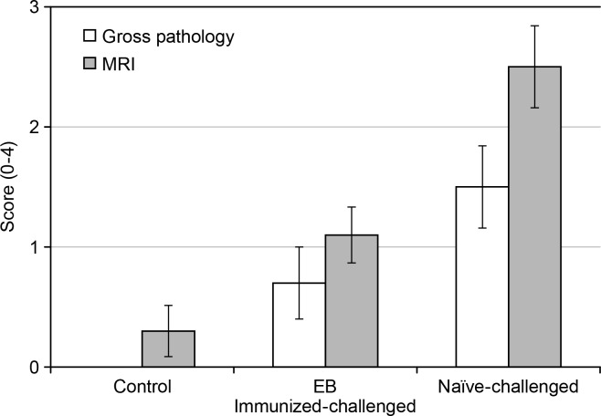
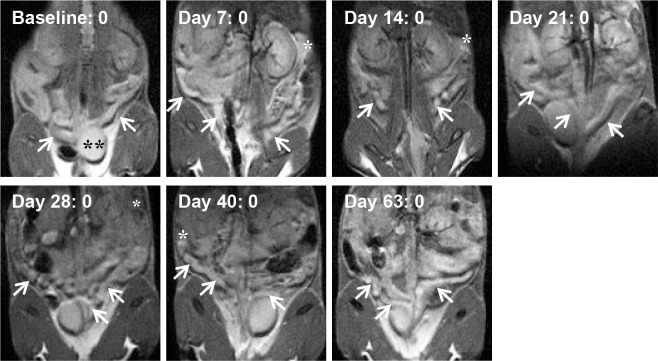
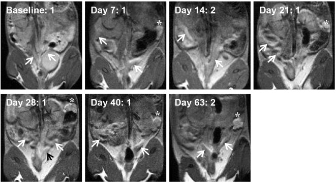
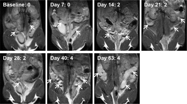
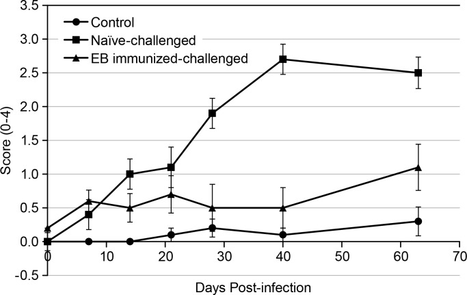
Chlamydia trachomatis is among the most prevalent of sexually transmitted diseases. While Chlamydia infection is a reportable event and screening has increased over time, enhanced surveillance has not resulted in a reduction in the rate of infections, and Chlamydia infections frequently recur. The development of a preventative vaccine for Chlamydia may be the only effective approach for reducing infection and the frequency of pathological outcomes. Current vaccine research efforts involve time consuming and/or invasive approaches for assessment of disease state, and MRI presents a clinically translatable method for assessing infection and related pathology both quickly and non-invasively. Longitudinal T2-weighted MRI was performed over 63 days on both control or Chlamydia muridarum challenged mice, either with or without elementary body (EB) immunization, and gross necropsy was performed on day 65. A scoring system was developed to assess the number of regions affected by Chlamydia pathology and was used to document pathology over time and at necropsy. The scoring system documented increasing incidence of pathology in the unimmunized and challenged mice (significantly greater compared to the control and EB immunized-challenged groups) by 21 days post-challenge. No differences between the unchallenged and EB immunized-challenged mice were observed. MRI scores at Day 63 were consistently higher than gross necropsy scores at Day 65, although two of the three groups of mice showed no significant differences between the two techniques. In this work we describe the application of MRI in mice for the potential evaluation of disease pathology and sequelae caused by C. muridarum infection and this technique's potential for evaluation of vaccines for Chlamydia.
沙眼衣原体是最常见的性传播疾病之一。虽然衣原体感染是应报告的事件,且随着时间推移筛查有所增加,但加强监测并未导致感染率下降,而且衣原体感染经常复发。开发一种针对衣原体的预防性疫苗可能是降低感染率和病理结果发生率的唯一有效方法。目前的疫苗研究工作涉及耗时和/或侵入性的疾病状态评估方法,而磁共振成像(MRI)提供了一种可在临床应用的方法,能够快速且非侵入性地评估感染及相关病理情况。对对照小鼠或感染鼠沙眼衣原体的小鼠进行了为期63天的纵向T2加权MRI检查,这些小鼠有的接种了原体(EB),有的未接种,在第65天进行大体尸检。开发了一种评分系统来评估受衣原体病理影响的区域数量,并用于记录随时间变化以及尸检时的病理情况。该评分系统记录了未免疫且受感染小鼠中病理发生率的增加(与对照组和接种EB并受感染的组相比显著更高),在感染后21天即可观察到。未受感染小鼠与接种EB并受感染的小鼠之间未观察到差异。第63天的MRI评分始终高于第65天的大体尸检评分,尽管三组小鼠中有两组在这两种技术之间未显示出显著差异。在这项工作中,我们描述了MRI在小鼠中的应用,用于潜在评估鼠沙眼衣原体感染引起的疾病病理和后遗症,以及该技术在评估衣原体疫苗方面的潜力。