Xie Yangli, Yi Lingxian, Weng Tujun, Huang Junlan, Luo Fengtao, Jiang Wanling, Xian Cory J, Du Xiaolan, Chen Lin
1. Center of Bone Metabolism and Repair, Department of Rehabilitation Medicine, State Key Laboratory of Trauma, Burns and Combined injury, Trauma Center, Research Institute of Surgery, Daping Hospital, Third Military Medical University, Chongqing 400042, China;
1. Center of Bone Metabolism and Repair, Department of Rehabilitation Medicine, State Key Laboratory of Trauma, Burns and Combined injury, Trauma Center, Research Institute of Surgery, Daping Hospital, Third Military Medical University, Chongqing 400042, China;; 3. Intensive Care Unit, The 306th hospital of PLA, Beijing 100101, China.
Int J Biol Sci. 2016 Jul 17;12(8):990-9. doi: 10.7150/ijbs.14077. eCollection 2016.
PTH stimulates bone formation in Fgfr3 knockout mice through promotion of proliferation and differentiation in osteoblasts.
Previous studies showed that endogenous fibroblast growth factor 2 (FGF-2) is required for parathyroid hormone (PTH)-stimulated bone anabolic effects, however, the exact mechanisms by which PTH stimulate bone formation and the function of FGF receptors in mediating these actions are not fully defined. FGF receptor 3 (FGFR3) has been characterized as an important regulator of bone metabolism and is confirmed to cross-talk with PTH/PTHrP signal in cartilage and bone development.
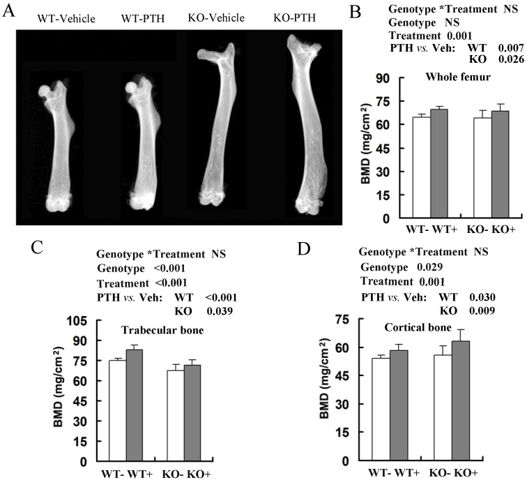
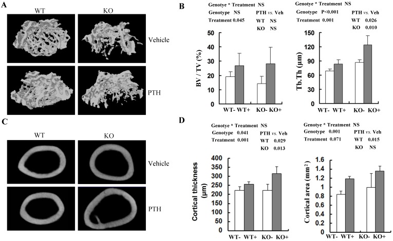
Fgfr3 knockout and wild-type mice at 2-month-old and 4-month-old were intraperitoneally injected with PTH intermittently for 4 weeks and then the skeletal responses to PTH were assessed by dual energy X-ray absorptiometry (DEXA), micro-computed tomography (μCT) and bone histomorphometry.
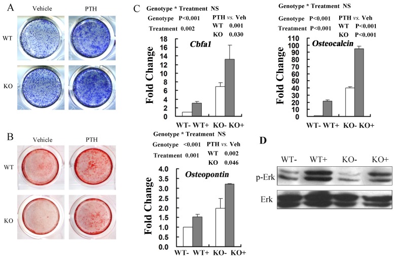
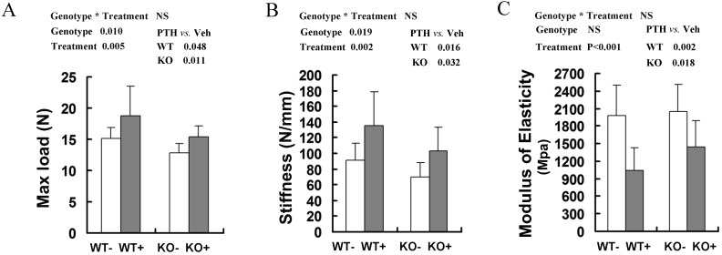
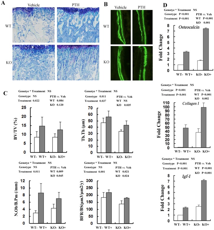
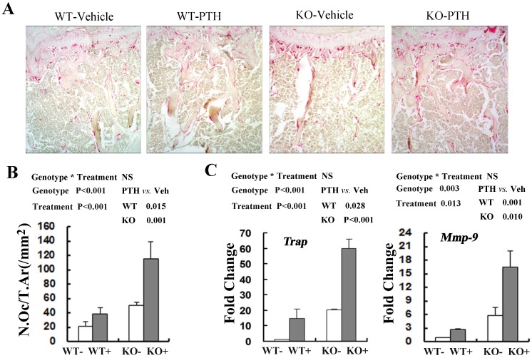
Intermittent PTH treatment improved bone mineral density (BMD) and femoral mechanical properties in both Fgfr3 (-/-) and wild-type mice. Histomorphometric analysis showed that bone formation and bone resorption were increased in both genotypes following PTH treatment. PTH treatment increased trabecular bone volume (BV/TV) in WT and Fgfr3-deficient mice. The anabolic response in Fgfr3-deficient and wild-type bone is characterized by an increase of both bone formation and resorption-related genes following PTH treatment. In addition, we found that Fgfr3 null osteoblasts (compared to wild-type controls) maintained normal abilities to response to PTH-stimulated increase of proliferation, differentiation, expression of osteoblastic marker genes (Cbfa1, Osteopontin and Osteocalcin), and phosphorylation of Erk1/2.
Bone anabolic effects of PTH were not impaired by the absence of FGFR3, suggesting that the FGFR3 signaling may not be required for osteoanabolic effects of PTH activities.
甲状旁腺激素(PTH)通过促进成骨细胞的增殖和分化,刺激Fgfr3基因敲除小鼠的骨形成。
先前的研究表明,内源性成纤维细胞生长因子2(FGF - 2)是甲状旁腺激素(PTH)刺激骨合成代谢作用所必需的,然而,PTH刺激骨形成的确切机制以及FGF受体在介导这些作用中的功能尚未完全明确。成纤维细胞生长因子3(FGFR3)已被确定为骨代谢的重要调节因子,并已证实在软骨和骨发育过程中与PTH/PTHrP信号相互作用。
对2月龄和4月龄的Fgfr3基因敲除小鼠和野生型小鼠进行为期4周的间歇性腹腔注射PTH,然后通过双能X线吸收法(DEXA)、显微计算机断层扫描(μCT)和骨组织形态计量学评估骨骼对PTH的反应。
间歇性PTH治疗改善了Fgfr3(-/-)和野生型小鼠的骨矿物质密度(BMD)和股骨力学性能。组织形态计量学分析表明,PTH治疗后两种基因型的骨形成和骨吸收均增加。PTH治疗增加了野生型和Fgfr3基因缺陷小鼠的小梁骨体积(BV/TV)。Fgfr3基因缺陷和野生型骨的合成代谢反应的特征是PTH治疗后骨形成和与吸收相关的基因均增加。此外,我们发现Fgfr3基因缺失的成骨细胞(与野生型对照相比)在对PTH刺激的增殖增加、分化、成骨细胞标记基因(Cbfa1、骨桥蛋白和骨钙素)表达以及Erk1/2磷酸化的反应中保持正常能力。
FGFR3的缺失并未损害PTH的骨合成代谢作用,这表明FGFR3信号传导可能不是PTH活性的骨合成代谢作用所必需的。