Choi Seung Ah, Kwak Pil Ae, Kim Seung-Ki, Park Sung-Hye, Lee Ji Yeoun, Wang Kyu-Chang, Oh Hyun Jeong, Kim Kyuwan, Lee Dong Soo, Hwang Do Won, Phi Ji Hoon
Division of Pediatric Neurosurgery, Seoul National University Children's Hospital, 101 Daehakro, Jongno-gu, 110-744, Seoul, Republic of Korea.
Department of Pathology, Seoul National University Hospital, College of Medicine, Seoul, Korea.
BMC Cancer. 2016 Sep 8;16(1):723. doi: 10.1186/s12885-016-2742-y.
The primary cause of treatment failure in medulloblastomas (MB) is the development of leptomeningeal dissemination (seeding). For translational research on MB seeding, one of the major challenges is the development of reliable experimental models that simulate the seeding and growth characteristics of MBs. To overcome this obstacle, we improved an experimental mouse model by intracisternal inoculation of human MB cells and monitoring with in vivo live images.
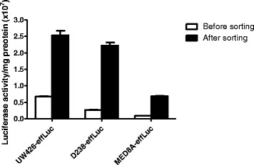
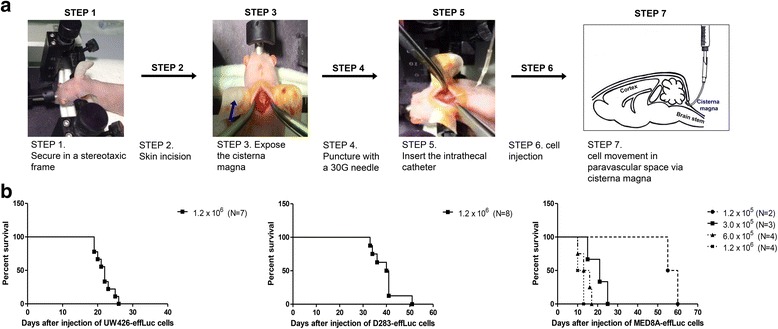
Human MB cells (UW426, D283 and MED8A) were transfected with a firefly luciferase gene and a Thy1.1 (CD90.1) marker linked with IRES under the control of the CMV promoter in a retroviral DNA backbone (effLuc). The MB-effLuc cells were injected into the cisterna magna using an intrathecal catheter, and bioluminescence images were captured. We performed histopathological analysis to confirm the extent of tumor seeding.
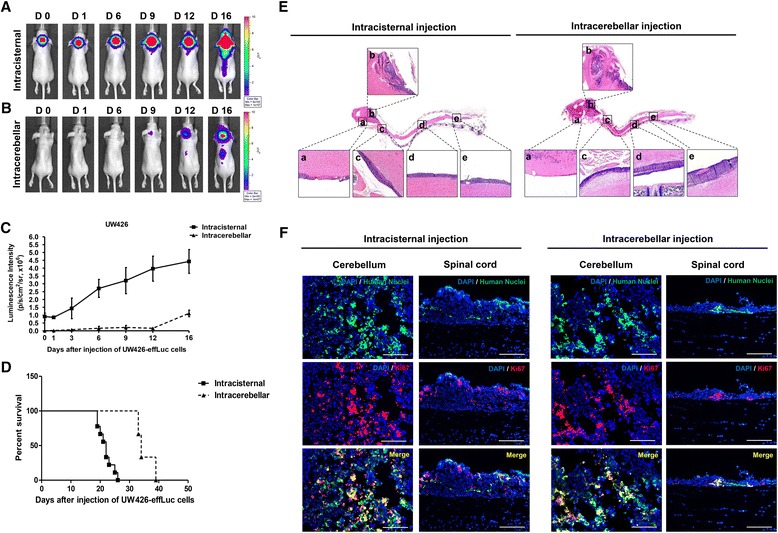
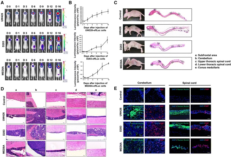
The luciferase activity of MB-effLuc cells displayed a gradually increasing pattern, which correlated with a quantitative luminometric assay. Live imaging showed that the MB-effLuc cells were diffusely distributed in the cervical spinal cord and the lumbosacral area. All mice injected with UW426-effLuc, D283-effLuc and MED8A-effLuc died within 51 days. The median survival was 22, 41 and 12 days after injection of 1.2 × 10(6) UW426-effLuc, D283-effLuc and MED8A-effLuc cells, respectively. The histopathological studies revealed that the MB-effLuc cells spread extensively and diffusely along the leptomeninges of the brain and spinal cord, forming tumor cell-coated layers. The tumor cells in the subarachnoid space expressed a human nuclei marker and Ki-67. Compared with the intracerebellar injection method in which the subfrontal area and distal spinal cord were spared by tumor cell seeding in some mice, the intracisternal injection model more closely resembled the widespread leptomeningeal seeding observed in MB patients.
The results and described method are valuable resources for further translational research to overcome MB seeding.
髓母细胞瘤(MB)治疗失败的主要原因是软脑膜播散(种植)的发生。对于MB种植的转化研究,主要挑战之一是开发能够模拟MB种植和生长特征的可靠实验模型。为克服这一障碍,我们通过脑池内接种人MB细胞并进行体内实时成像监测,改进了一种实验小鼠模型。
将萤火虫荧光素酶基因和与内部核糖体进入位点(IRES)相连的Thy1.1(CD90.1)标记物在巨细胞病毒(CMV)启动子控制下,转染到逆转录病毒DNA骨架(effLuc)中的人MB细胞(UW426、D283和MED8A)。使用鞘内导管将MB-effLuc细胞注入小脑延髓池,并采集生物发光图像。我们进行了组织病理学分析以确认肿瘤种植的范围。
MB-effLuc细胞的荧光素酶活性呈逐渐增加的模式,这与定量发光测定相关。实时成像显示MB-effLuc细胞广泛分布于颈髓和腰骶部区域。所有注射UW426-effLuc、D283-effLuc和MED8A-effLuc的小鼠均在51天内死亡。分别注射1.2×10⁶ UW426-effLuc、D283-effLuc和MED8A-effLuc细胞后,中位生存期分别为22天、41天和12天。组织病理学研究显示,MB-effLuc细胞沿脑和脊髓的软脑膜广泛弥漫性扩散,形成肿瘤细胞包被层。蛛网膜下腔的肿瘤细胞表达人核标记物和Ki-67。与小脑内注射方法相比,在小脑内注射方法中部分小鼠的额下区域和脊髓远端未出现肿瘤细胞种植,而脑池内注射模型更类似于在MB患者中观察到的广泛软脑膜种植。
这些结果和所描述的方法是进一步开展克服MB种植的转化研究的宝贵资源。