Dasgupta Swapan K, Le Anhquyen, Da Qi, Cruz Miguel, Rumbaut Rolando E, Thiagarajan Perumal
Department of Pathology and Immunology, Baylor College of Medicine, Houston, Texas, United States of America.
Center for Translational Research on Inflammatory Diseases (CTRID), Michael E. DeBakey Veterans Affairs Medical Center, Houston, Texas, United States of America.
PLoS One. 2016 Sep 14;11(9):e0162897. doi: 10.1371/journal.pone.0162897. eCollection 2016.
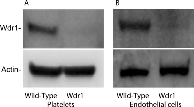
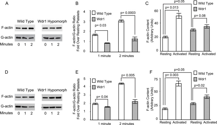
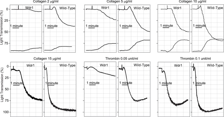
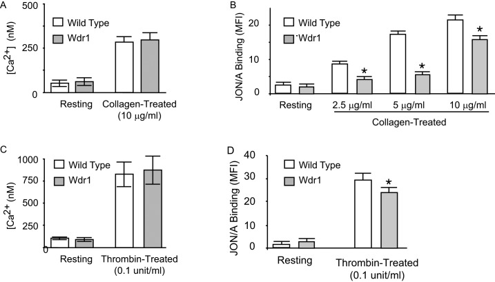
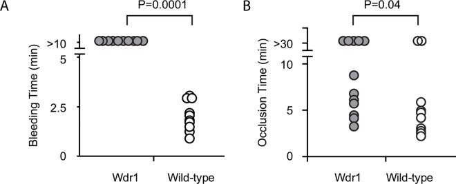
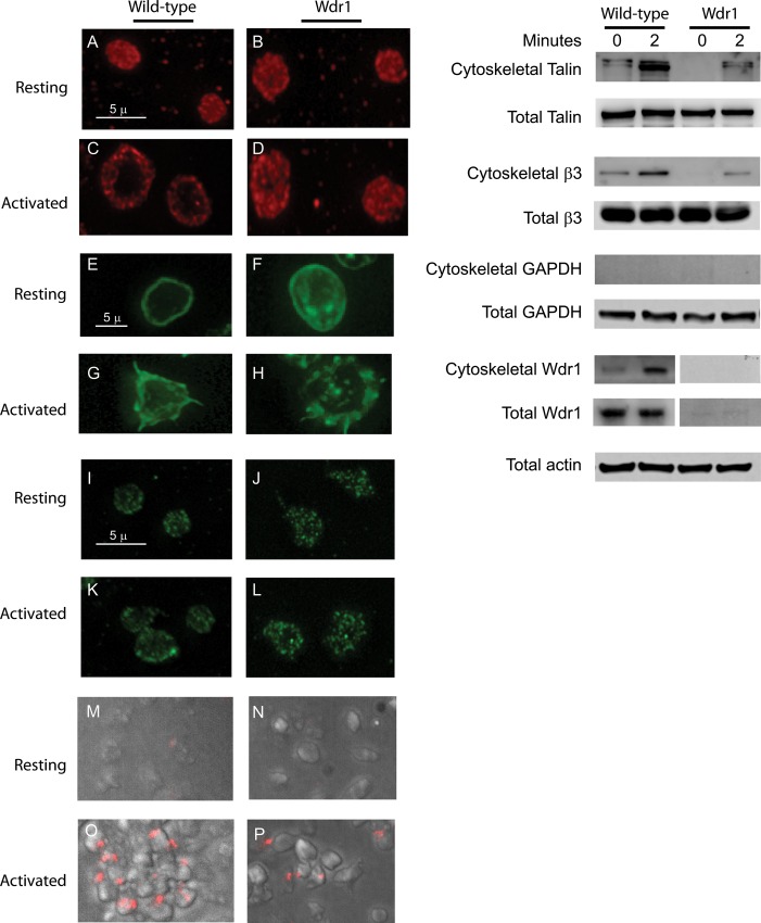
In resting platelets, the integrin αIIbβ3 is present in a low-affinity "bent" state. During platelet aggregation, intracytoplasmic signals induce conformational changes (inside-out signaling) that result in a "swung-out" conformation competent to bind ligands such as fibrinogen. The cytoskeleton plays an essential role in αIIbβ3 activation. We investigated the role of the actin interacting protein Wdr1 in αIIbβ3 activation. Wdr1-hypomorphic mice had a prolonged bleeding time (> 10 minutes) compared to that of wild-type mice (2.1 ± 0.7 minutes). Their platelets had impaired aggregation to collagen and thrombin. In a FeCl3 induced carotid artery thrombosis model, vessel occlusion in Wdr1-hypomorphic mice was prolonged significantly compared to wild-type mice (9.0 ± 10.5 minutes versus 5.8 ± 12.6 minutes (p = 0.041). Activation-induced binding of JON/A (a conformation-specific antibody to activated αIIbβ3) was significantly less in Wdr1-hypomorphic platelets at various concentrations of collagen, indicating impaired inside-out activation of αIIbβ3, despite a normal calcium response. Actin turnover, assessed by measuring F-actin and G-actin ratios during collagen- and thrombin-induced platelet aggregation, was highly impaired in Wdr1-hypomorphic platelets. Furthermore, talin failed to redistribute and translocate to the cytoskeleton following activation in Wdr1-hypomorphic platelets. These studies show that Wdr1 is essential for talin-induced activation of αIIbβ3 during platelet activation.
在静息血小板中,整合素αIIbβ3以低亲和力的“弯曲”状态存在。在血小板聚集过程中,胞浆内信号诱导构象变化(由内向外信号传导),导致形成一种能够结合纤维蛋白原等配体的“伸展”构象。细胞骨架在αIIbβ3激活中起重要作用。我们研究了肌动蛋白相互作用蛋白Wdr1在αIIbβ3激活中的作用。与野生型小鼠(2.1±0.7分钟)相比,Wdr1低表达小鼠的出血时间延长(>10分钟)。它们的血小板对胶原蛋白和凝血酶的聚集能力受损。在FeCl3诱导的颈动脉血栓形成模型中,与野生型小鼠相比,Wdr1低表达小鼠的血管阻塞时间显著延长(9.0±10.5分钟对5.8±12.6分钟(p = 0.041))。在不同浓度的胶原蛋白作用下,Wdr1低表达血小板中JON/A(一种针对活化αIIbβ3的构象特异性抗体)的激活诱导结合显著减少,这表明尽管钙反应正常,但αIIbβ3的由内向外激活受损。通过测量胶原蛋白和凝血酶诱导的血小板聚集过程中F-肌动蛋白和G-肌动蛋白的比例来评估的肌动蛋白周转,在Wdr1低表达血小板中严重受损。此外,在Wdr1低表达血小板激活后,踝蛋白未能重新分布并转运至细胞骨架。这些研究表明,Wdr1在血小板激活过程中对踝蛋白诱导的αIIbβ3激活至关重要。