Vähätupa Maria, Prince Stuart, Vataja Suvi, Mertimo Teija, Kataja Marko, Kinnunen Kati, Marjomäki Varpu, Uusitalo Hannu, Komatsu Masanobu, Järvinen Tero A H, Uusitalo-Järvinen Hannele
Department of Ophthalmology, University of Tampere, Tampere, Finland 2Department of Anatomy, University of Tampere, Tampere, Finland.
Department of Anatomy, University of Tampere, Tampere, Finland.
Invest Ophthalmol Vis Sci. 2016 Sep 1;57(11):4898-4909. doi: 10.1167/iovs.16-19212.
The role of R-Ras in retinal angiogenesis and vascular permeability was evaluated in an oxygen-induced retinopathy (OIR) model using R-Ras knockout (KO) mice and in human diabetic neovascular membranes.
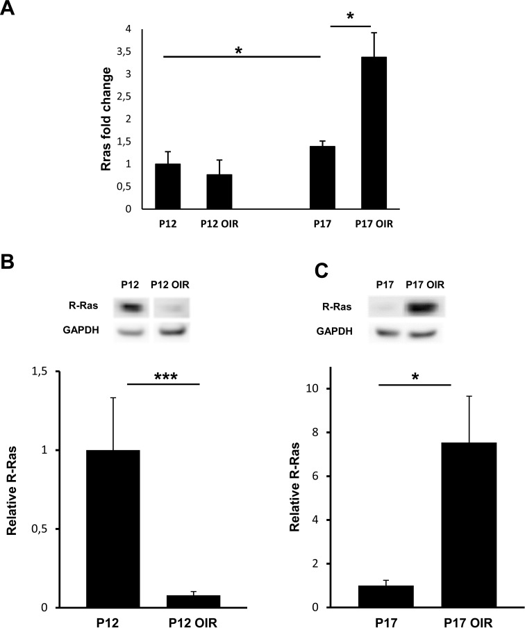
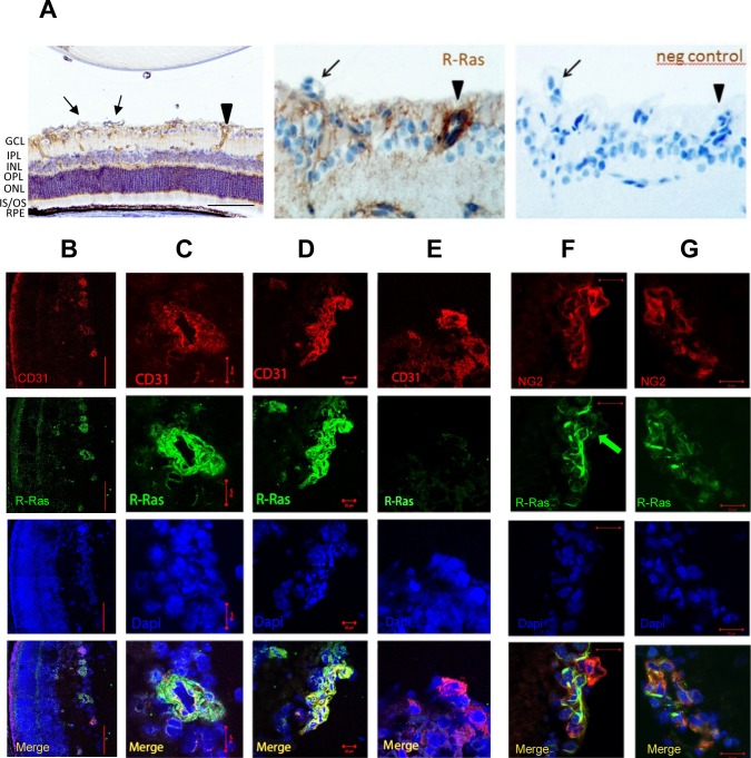
Mice deficient for R-Ras and their wild-type (WT) littermates were subjected to 75% oxygen from postnatal day 7 (P7) to P12 and then returned to room air. At P17 retinal vascularization was examined from whole mounts, and retinal vascular permeability was studied using Miles assay. Real-time RT-PCR, Western blotting, and immunohistochemistry were used to assess the expression of R-Ras in retina during development or in the OIR model. The degree of pericyte coverage and vascular endothelial (VE)-cadherin expression on WT and R-Ras KO retinal blood vessels was quantified using confocal microscopy. The correlation of R-Ras with vascular endothelial growth factor receptor 2 (VEGFR2) and human serum albumin on human proliferative diabetic retinopathy membranes was assessed using immunohistochemistry.
In retina, R-Ras expression was mostly restricted to the vasculature. Retinal vessels in the R-Ras KO mice were significantly more permeable than WT controls in the OIR model. A significant reduction in the direct physical contact between pericytes and blood vessel endothelium as well as reduced VE-cadherin immunostaining was found in R-Ras-deficient mice. In human proliferative diabetic retinopathy neovascular membranes, R-Ras expression negatively correlated with increased vascular leakage and expression of VEGFR2, a marker of blood vessel immaturity.
Our results suggest that R-Ras has a role in controlling retinal vessel maturation and stabilization in ischemic retinopathy and provides a potential target for pharmacologic manipulation to treat diabetic retinopathy.
利用R-Ras基因敲除(KO)小鼠在氧诱导视网膜病变(OIR)模型中以及在人类糖尿病新生血管膜中评估R-Ras在视网膜血管生成和血管通透性中的作用。
R-Ras基因缺陷小鼠及其野生型(WT)同窝小鼠在出生后第7天(P7)至P12接受75%的氧气处理,然后放回正常空气中。在P17时,从视网膜全层标本检查视网膜血管生成情况,并使用迈尔斯试验研究视网膜血管通透性。采用实时逆转录聚合酶链反应(RT-PCR)、蛋白质印迹法和免疫组织化学法评估R-Ras在视网膜发育过程中或OIR模型中的表达。使用共聚焦显微镜对WT和R-Ras KO视网膜血管上的周细胞覆盖程度和血管内皮(VE)-钙黏蛋白表达进行定量分析。采用免疫组织化学法评估R-Ras与人类增殖性糖尿病视网膜病变膜上血管内皮生长因子受体2(VEGFR2)和人血清白蛋白的相关性。
在视网膜中,R-Ras表达主要局限于血管系统。在OIR模型中,R-Ras KO小鼠的视网膜血管通透性明显高于WT对照组。在R-Ras基因缺陷小鼠中,发现周细胞与血管内皮之间的直接物理接触显著减少,且VE-钙黏蛋白免疫染色降低。在人类增殖性糖尿病视网膜病变新生血管膜中,R-Ras表达与血管渗漏增加以及血管不成熟标志物VEGFR2的表达呈负相关。
我们的结果表明,R-Ras在缺血性视网膜病变中对控制视网膜血管成熟和稳定起作用,并为治疗糖尿病视网膜病变的药物干预提供了一个潜在靶点。