Pang Paul, Abbott Molly, Chang Steven L, Abdi Malyun, Chauhan Nikita, Mistri Murti, Ghofrani Joshua, Fucci Quynh-Anh, Walker Colleen, Leonardi Corey, Grady Samuel, Halim Arvin, Hoffman Ryan, Lu Tzongshi, Cao Huixia, Tullius Stefan G, Malek Sayeed, Kumar Sanjaya, Steele Graeme, Kibel Adam, Freedman Benjamin S, Waikar Sushrut S, Siedlecki Andrew M
Department of Internal Medicine, Brigham and Women's Hospital, Harvard Medical School, Boston, Massachusetts, USA.
Urology Division, Department of Surgery, Brigham and Women's Hospital, Harvard Medical School, Boston, Massachusetts, USA.
Kidney Int. 2017 Jan;91(1):129-143. doi: 10.1016/j.kint.2016.07.037. Epub 2016 Sep 29.
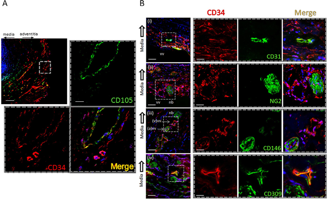
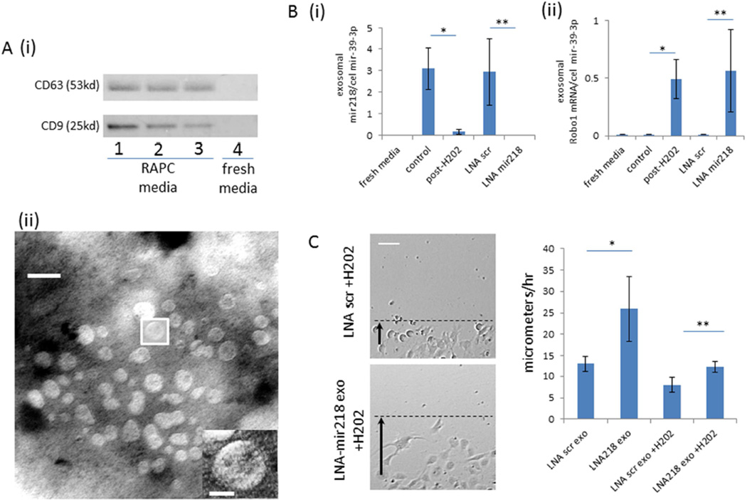
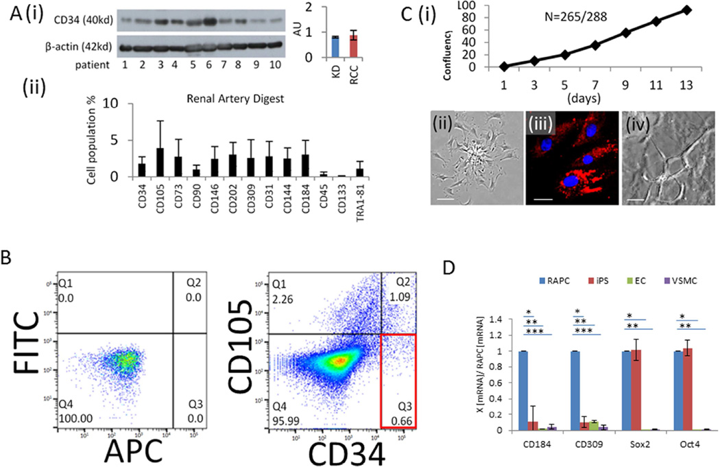
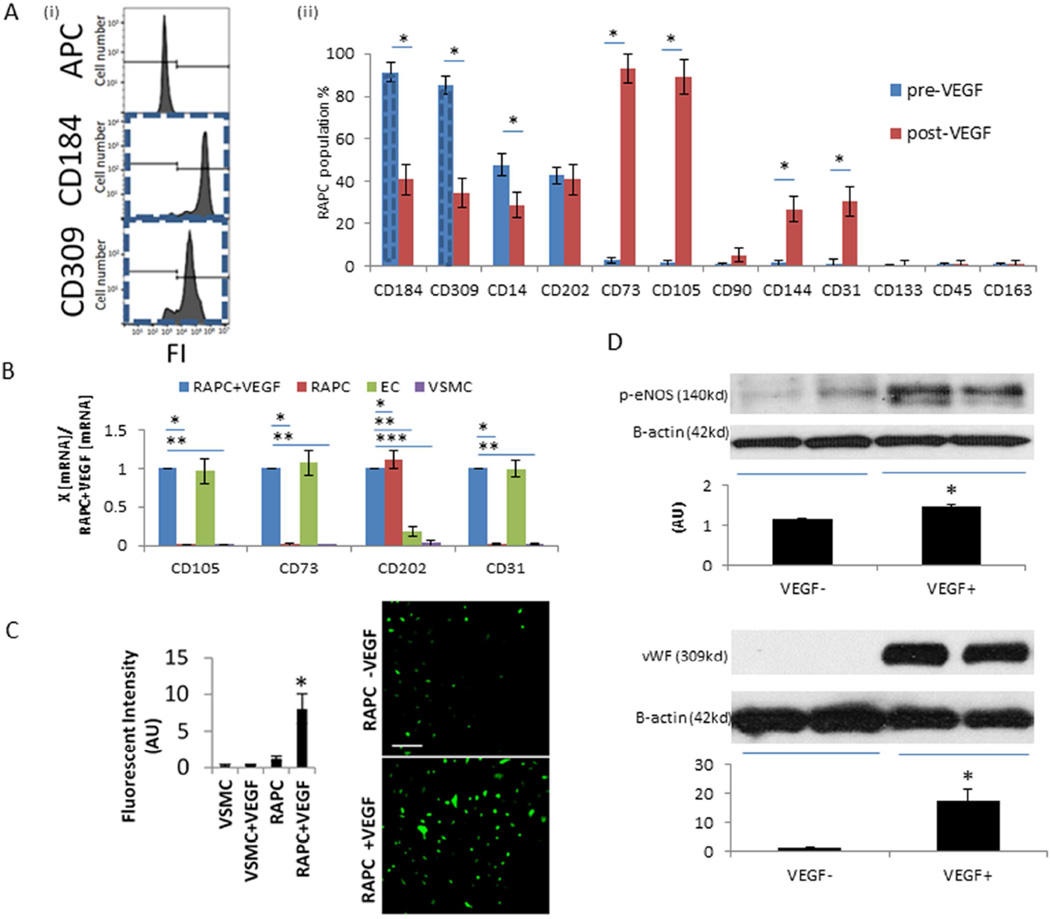
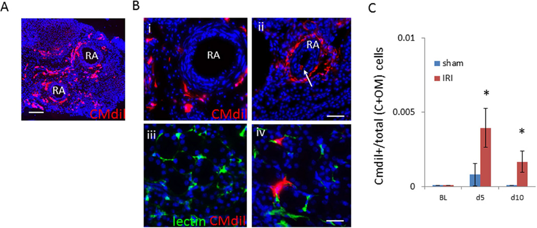
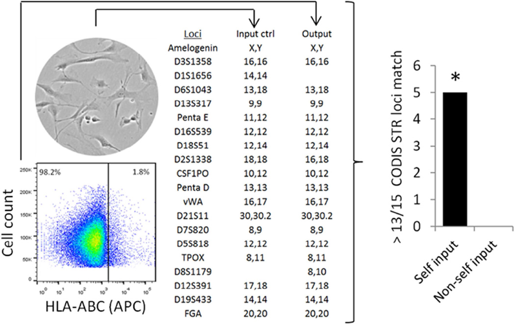
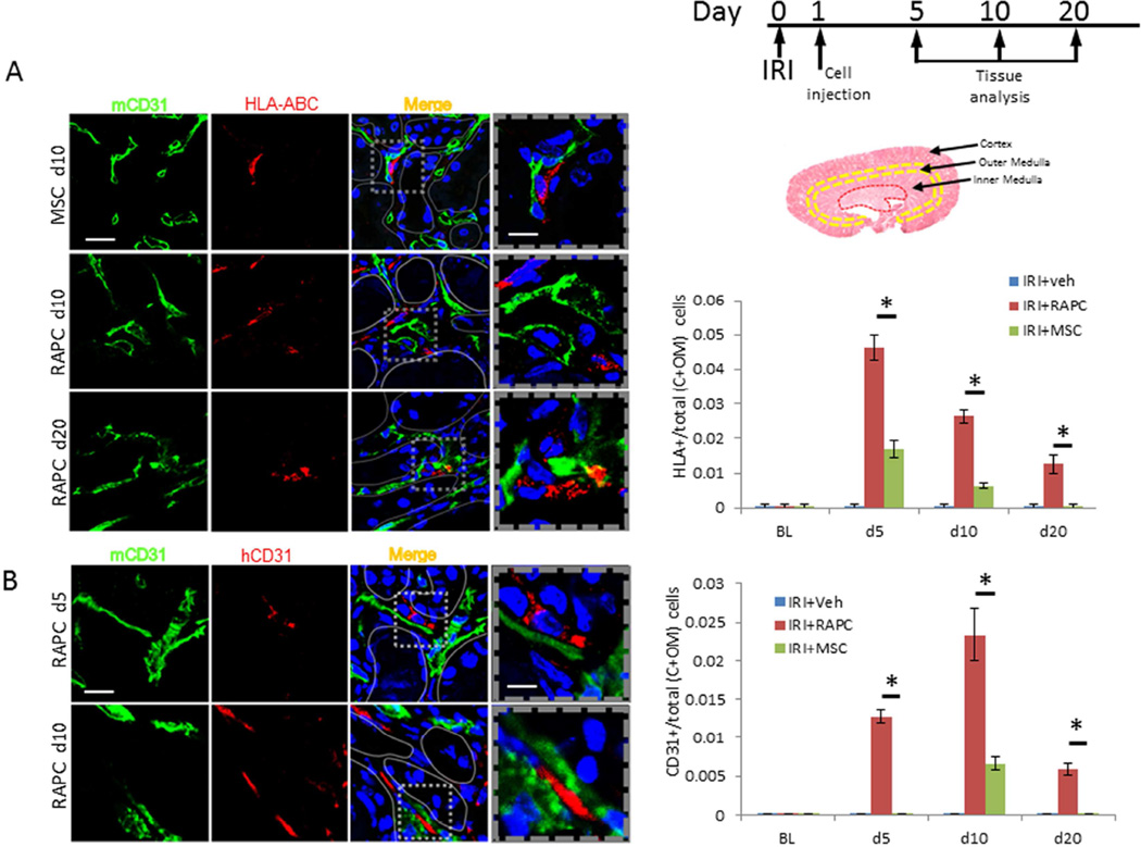
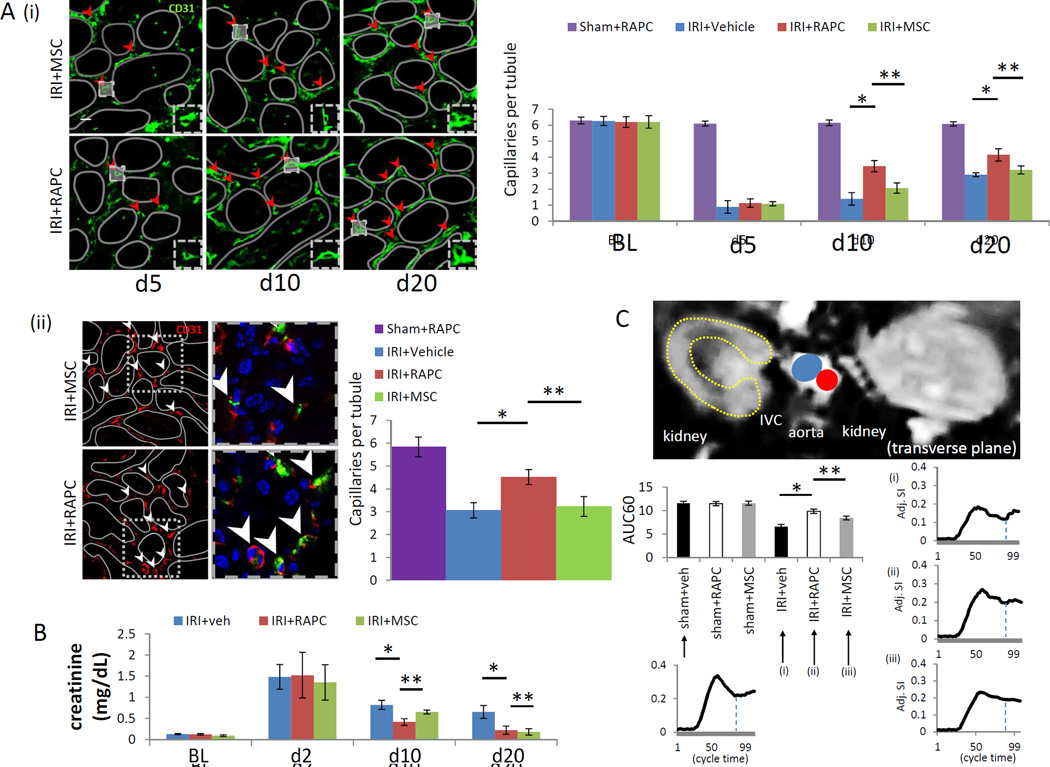
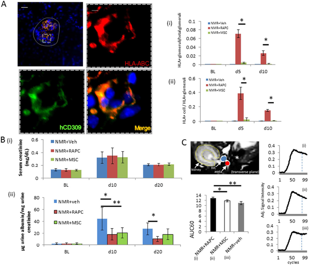
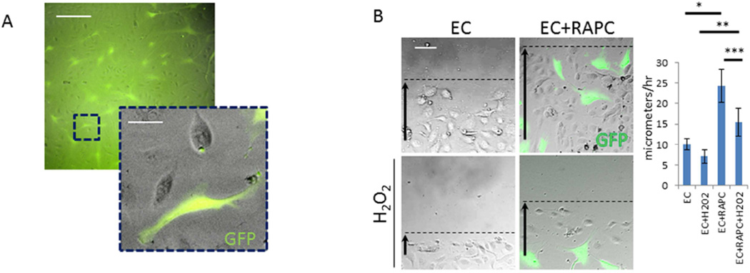
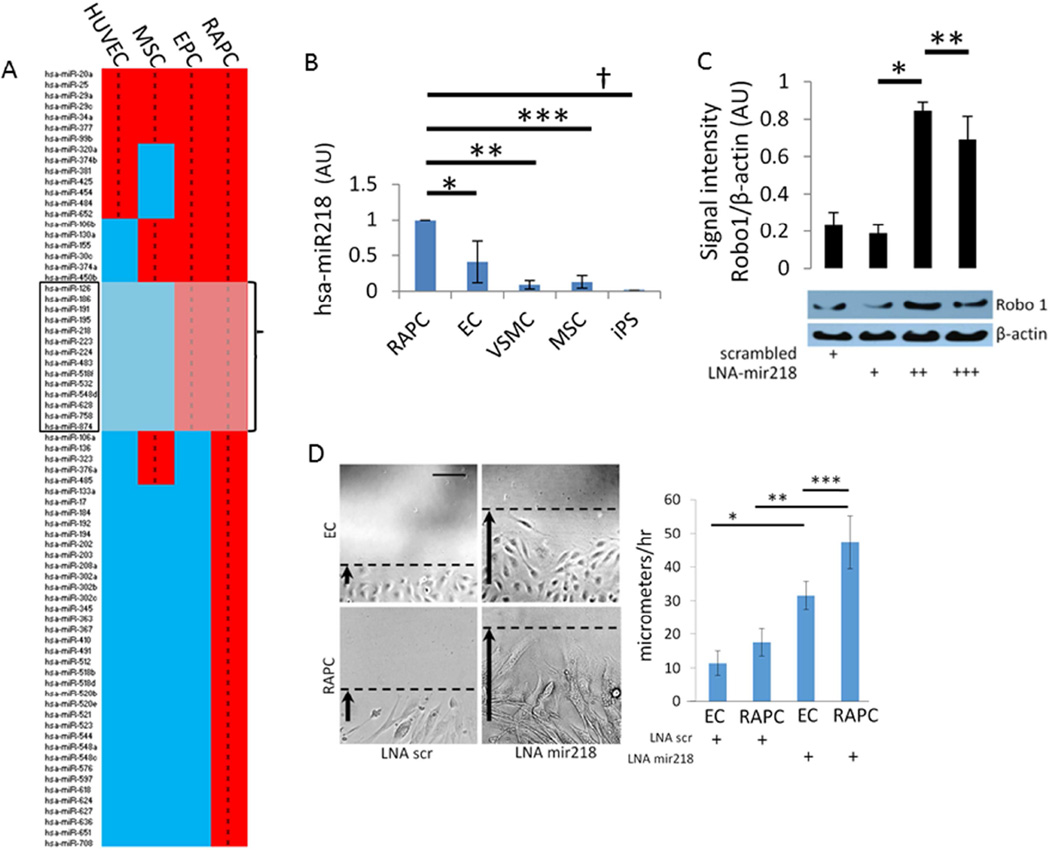
Vascular progenitor cells show promise for the treatment of microvasculature endothelial injury. We investigated the function of renal artery progenitor cells derived from radical nephrectomy patients, in animal models of acute ischemic and hyperperfusion injuries. Present in human adventitia, CD34positive/CD105negative cells were clonal and expressed transcription factors Sox2/Oct4 as well as surface markers CXCR4 (CD184)/KDR(CD309) consistent with endothelial progenitor cells. Termed renal artery-derived vascular progenitor cells (RAPC), injected cells were associated with decreased serum creatinine after ischemia/reperfusion, reduced albuminuria after hyperperfusion, and improved blood flow in both models. A small population of RAPC integrated with the renal microvasculature following either experimental injury. At a cellular level, RAPC promoted local endothelial migration in co-culture. Profiling of RAPC microRNA identified high levels of miRNA 218; also found at high levels in exosomes isolated from RAPC conditioned media after cell contact for 24 hours. After hydrogen peroxide-induced endothelial injury, RAPC exosomes harbored Robo-1 transcript; a gene known to be regulated by mir218. Such exosomes enhanced endothelial cell migration in culture in the absence of RAPC. Thus, our work shows the feasibility of pre-emptive pro-angiogenic progenitor cell procurement from a targeted patient population and potential therapeutic use in the form of autologous cell transplantation.
血管祖细胞在治疗微血管内皮损伤方面显示出前景。我们在急性缺血和高灌注损伤的动物模型中,研究了来自根治性肾切除术患者的肾动脉祖细胞的功能。存在于人类外膜中的CD34阳性/CD105阴性细胞具有克隆性,表达转录因子Sox2/Oct4以及与内皮祖细胞一致的表面标志物CXCR4(CD184)/KDR(CD309)。这些细胞被称为肾动脉来源的血管祖细胞(RAPC),注射的细胞与缺血/再灌注后血清肌酐降低、高灌注后蛋白尿减少以及两种模型中的血流改善有关。在两种实验性损伤后,一小部分RAPC与肾微血管整合。在细胞水平上,RAPC在共培养中促进局部内皮细胞迁移。对RAPC微小RNA的分析发现miRNA 218水平较高;在细胞接触24小时后从RAPC条件培养基中分离的外泌体中也发现其水平较高。在过氧化氢诱导的内皮损伤后,RAPC外泌体含有Robo-1转录本;这是一个已知受mir218调控的基因。这种外泌体在没有RAPC的情况下增强了培养中的内皮细胞迁移。因此,我们的工作表明从目标患者群体中预先获取促血管生成祖细胞的可行性以及自体细胞移植形式的潜在治疗用途。