• 文献检索
  • 文档翻译
  • 深度研究
  • 学术资讯
  • Suppr Zotero 插件Zotero 插件
  • 邀请有礼
  • 套餐&价格
  • 历史记录
应用&插件
Suppr Zotero 插件Zotero 插件浏览器插件Mac 客户端Windows 客户端微信小程序
定价
高级版会员购买积分包购买API积分包
服务
文献检索文档翻译深度研究API 文档MCP 服务
关于我们
关于 Suppr公司介绍联系我们用户协议隐私条款
关注我们

Suppr 超能文献

核心技术专利:CN118964589B侵权必究
粤ICP备2023148730 号-1Suppr @ 2026

文献检索

告别复杂PubMed语法,用中文像聊天一样搜索,搜遍4000万医学文献。AI智能推荐,让科研检索更轻松。

立即免费搜索

文件翻译

保留排版,准确专业,支持PDF/Word/PPT等文件格式,支持 12+语言互译。

免费翻译文档

深度研究

AI帮你快速写综述,25分钟生成高质量综述,智能提取关键信息,辅助科研写作。

立即免费体验

枯否细胞在肝内胆管癌的一种常见小鼠模型中诱导 Notch 介导的肝细胞转化。

Kupffer cells induce Notch-mediated hepatocyte conversion in a common mouse model of intrahepatic cholangiocarcinoma.

机构信息

Division of Organogenesis and Regeneration, Medical Institute of Bioregulation, Kyushu University, 3-1-1 Maidashi, Higashi-ku, Fukuoka 812-8582, Japan.

Division of Transcriptomics, Medical Institute of Bioregulation, Kyushu University, 3-1-1 Maidashi, Higashi-ku, Fukuoka 812-8582, Japan.

出版信息

Sci Rep. 2016 Oct 4;6:34691. doi: 10.1038/srep34691.

DOI:10.1038/srep34691
PMID:27698452
原文链接:https://pmc.ncbi.nlm.nih.gov/articles/PMC5048166/
Abstract

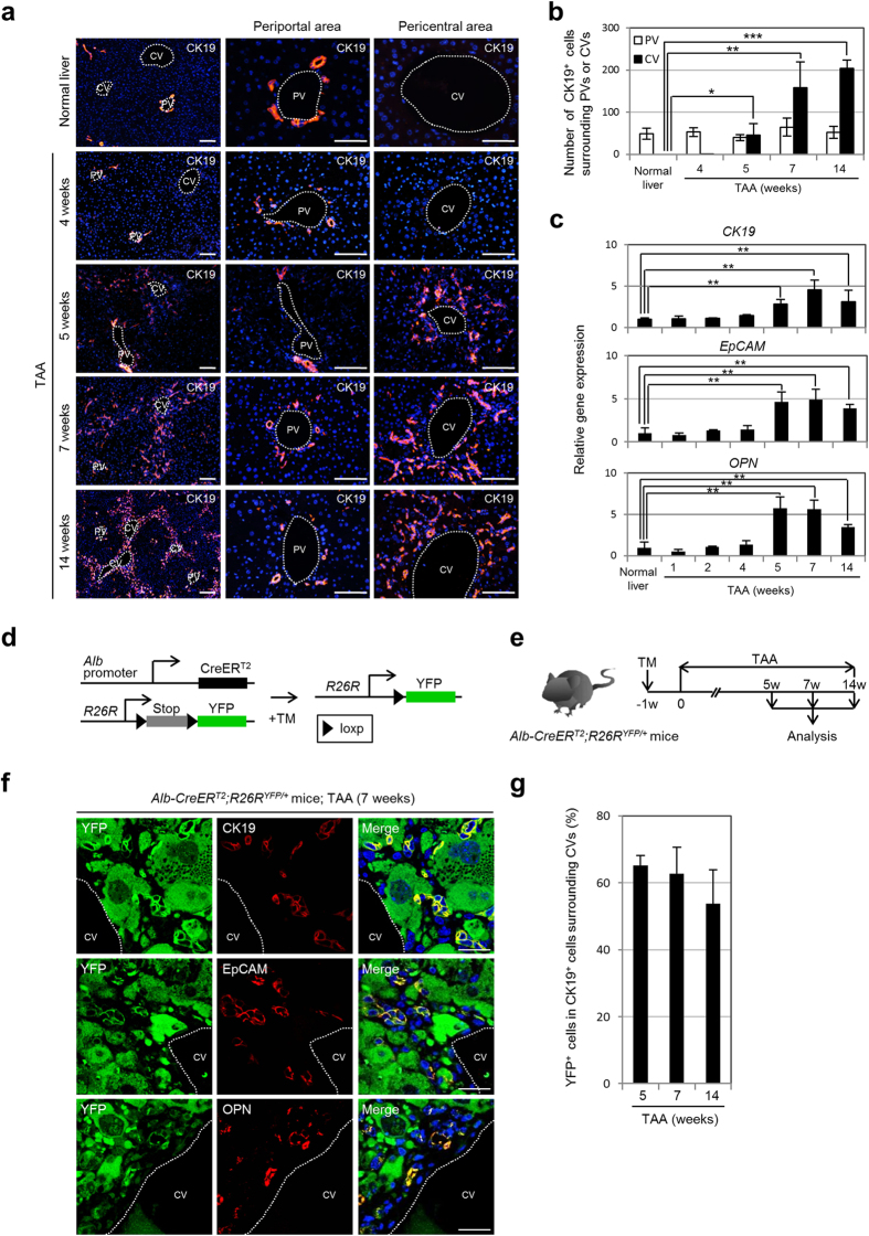
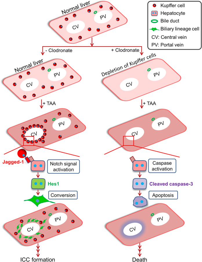
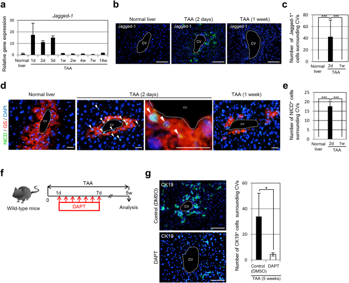
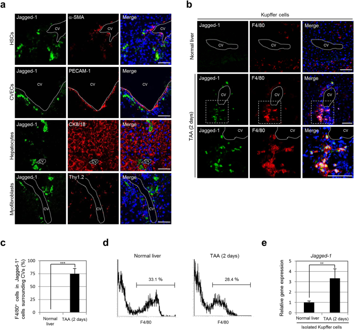
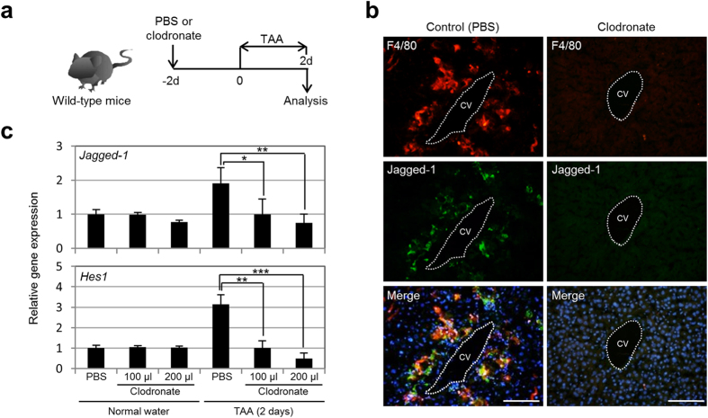
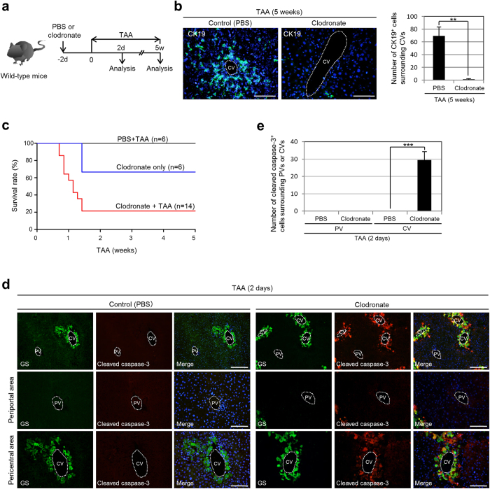
Intrahepatic cholangiocarcinoma (ICC) is a malignant epithelial neoplasm composed of cells resembling cholangiocytes that line the intrahepatic bile ducts in portal areas of the hepatic lobule. Although ICC has been defined as a tumor arising from cholangiocyte transformation, recent evidence from genetic lineage-tracing experiments has indicated that hepatocytes can be a cellular origin of ICC by directly changing their fate to that of biliary lineage cells. Notch signaling has been identified as an essential factor for hepatocyte conversion into biliary lineage cells at the onset of ICC. However, the mechanisms underlying Notch signal activation in hepatocytes remain unclear. Here, using a mouse model of ICC, we found that hepatic macrophages called Kupffer cells transiently congregate around the central veins in the liver and express the Notch ligand Jagged-1 coincident with Notch activation in pericentral hepatocytes. Depletion of Kupffer cells prevents the Notch-mediated cell-fate conversion of hepatocytes to biliary lineage cells, inducing hepatocyte apoptosis and increasing mortality in mice. These findings will be useful for uncovering the pathogenic mechanism of ICC and developing prevenient and therapeutic strategies for this refractory disease.

摘要

肝内胆管癌 (ICC) 是一种恶性上皮性肿瘤,由类似于肝小叶门脉区胆管内的胆管细胞的细胞组成。虽然 ICC 已被定义为源自胆管细胞转化的肿瘤,但最近的遗传谱系追踪实验证据表明,肝细胞可以通过直接改变其命运为胆管谱系细胞而成为 ICC 的细胞起源。 Notch 信号已被确定为 ICC 起始时肝细胞向胆管谱系细胞转化的必需因素。然而,Notch 信号在肝细胞中激活的机制尚不清楚。在这里,我们使用 ICC 的小鼠模型发现,称为库普弗细胞的肝巨噬细胞在肝脏中央静脉周围短暂聚集,并表达 Notch 配体 Jagged-1,同时在中央周围肝细胞中激活 Notch。耗尽库普弗细胞可防止 Notch 介导的肝细胞向胆管谱系细胞的命运转化,诱导肝细胞凋亡并增加小鼠的死亡率。这些发现将有助于揭示 ICC 的发病机制,并为这种难治性疾病开发预防和治疗策略。

https://cdn.ncbi.nlm.nih.gov/pmc/blobs/c803/5048166/82e325f31a27/srep34691-f6.jpg
https://cdn.ncbi.nlm.nih.gov/pmc/blobs/c803/5048166/e208f6a08452/srep34691-f1.jpg
https://cdn.ncbi.nlm.nih.gov/pmc/blobs/c803/5048166/0aa12a72a727/srep34691-f2.jpg
https://cdn.ncbi.nlm.nih.gov/pmc/blobs/c803/5048166/82372a606ad0/srep34691-f3.jpg
https://cdn.ncbi.nlm.nih.gov/pmc/blobs/c803/5048166/7b7df5ea472b/srep34691-f4.jpg
https://cdn.ncbi.nlm.nih.gov/pmc/blobs/c803/5048166/598f10162993/srep34691-f5.jpg
https://cdn.ncbi.nlm.nih.gov/pmc/blobs/c803/5048166/82e325f31a27/srep34691-f6.jpg

相似文献

1
Kupffer cells induce Notch-mediated hepatocyte conversion in a common mouse model of intrahepatic cholangiocarcinoma.枯否细胞在肝内胆管癌的一种常见小鼠模型中诱导 Notch 介导的肝细胞转化。
Sci Rep. 2016 Oct 4;6:34691. doi: 10.1038/srep34691.
2
Intrahepatic cholangiocarcinoma can arise from Notch-mediated conversion of hepatocytes.肝内胆管癌可源于 Notch 介导的肝细胞转化。
J Clin Invest. 2012 Nov;122(11):3914-8. doi: 10.1172/JCI63065.
3
Notch2 controls hepatocyte-derived cholangiocarcinoma formation in mice.Notch2 控制小鼠肝细胞源性胆管癌的形成。
Oncogene. 2018 Jun;37(24):3229-3242. doi: 10.1038/s41388-018-0188-1. Epub 2018 Mar 16.
4
Cholangiocarcinomas can originate from hepatocytes in mice.胆管癌可起源于小鼠的肝细胞。
J Clin Invest. 2012 Aug;122(8):2911-5. doi: 10.1172/JCI63212. Epub 2012 Jul 17.
5
Notch1 is overexpressed in human intrahepatic cholangiocarcinoma and is associated with its proliferation, invasiveness and sensitivity to 5-fluorouracil in vitro.Notch1 在人肝内胆管癌中过表达,并与肿瘤的增殖、侵袭以及对体外 5-氟尿嘧啶的敏感性相关。
Oncol Rep. 2014 Jun;31(6):2515-24. doi: 10.3892/or.2014.3123. Epub 2014 Apr 2.
6
Cell lineage tracing reveals a biliary origin of intrahepatic cholangiocarcinoma.细胞谱系追踪揭示了肝内胆管癌的胆管起源。
Cancer Res. 2014 Feb 15;74(4):1005-10. doi: 10.1158/0008-5472.CAN-13-1911. Epub 2013 Dec 5.
7
Is Essential in Proliferating Ductal Cell-Mediated Development of Intrahepatic Cholangiocarcinoma.在增殖性胆管细胞介导的肝内胆管癌发生发展中起关键作用。
Cancer Res. 2020 Dec 1;80(23):5305-5316. doi: 10.1158/0008-5472.CAN-20-1161. Epub 2020 Oct 16.
8
Epithelial Transforming Growth Factor-β Signaling Does Not Contribute to Liver Fibrosis but Protects Mice From Cholangiocarcinoma.上皮转化生长因子-β信号通路对肝纤维化无作用,但可保护小鼠免受胆管癌侵害。
Gastroenterology. 2016 Mar;150(3):720-33. doi: 10.1053/j.gastro.2015.11.039. Epub 2015 Nov 26.
9
Activation of Notch signaling is required for cholangiocarcinoma progression and is enhanced by inactivation of p53 in vivo.Notch 信号的激活对于胆管癌的进展是必需的,并且在体内 p53 的失活增强了其活性。
PLoS One. 2013 Oct 30;8(10):e77433. doi: 10.1371/journal.pone.0077433. eCollection 2013.
10
Notch1 Drives the Formation and Proliferation of Intrahepatic Cholangiocarcinoma.Notch1 驱动肝内胆管癌的形成和增殖。
Curr Med Sci. 2019 Dec;39(6):929-937. doi: 10.1007/s11596-019-2125-0. Epub 2019 Dec 16.

引用本文的文献

1
Diverse functions of SOX9 in liver development and homeostasis and hepatobiliary diseases.SOX9在肝脏发育、稳态及肝胆疾病中的多种功能。
Genes Dis. 2023 Jun 24;11(4):100996. doi: 10.1016/j.gendis.2023.03.035. eCollection 2024 Jul.
2
A Novel Mouse Model of Intrahepatic Cholangiocarcinoma Induced by Azoxymethane.一种由偶氮甲烷诱导的肝内胆管癌新型小鼠模型。
Int J Mol Sci. 2023 Sep 26;24(19):14581. doi: 10.3390/ijms241914581.
3
The crosstalk between parenchymal cells and macrophages: A keeper of tissue homeostasis.实质细胞与巨噬细胞的串扰:组织内稳态的维持者。

本文引用的文献

1
Hybrid Periportal Hepatocytes Regenerate the Injured Liver without Giving Rise to Cancer.混合性门周肝细胞可使受损肝脏再生而不引发癌症。
Cell. 2015 Aug 13;162(4):766-79. doi: 10.1016/j.cell.2015.07.026.
2
Self-renewing diploid Axin2(+) cells fuel homeostatic renewal of the liver.自我更新的二倍体Axin2(+)细胞为肝脏的稳态更新提供动力。
Nature. 2015 Aug 13;524(7564):180-5. doi: 10.1038/nature14863. Epub 2015 Aug 5.
3
Lineage fate of ductular reactions in liver injury and carcinogenesis.肝损伤和致癌过程中胆管反应的谱系命运
Front Immunol. 2022 Nov 24;13:1050188. doi: 10.3389/fimmu.2022.1050188. eCollection 2022.
4
Analysis of viral integration reveals new insights of oncogenic mechanism in HBV-infected intrahepatic cholangiocarcinoma and combined hepatocellular-cholangiocarcinoma.病毒整合分析揭示了乙型肝炎病毒感染的肝内胆管癌和肝细胞癌-胆管细胞癌合并癌中致癌机制的新见解。
Hepatol Int. 2022 Dec;16(6):1339-1352. doi: 10.1007/s12072-022-10419-3. Epub 2022 Sep 20.
5
Role of Immune Cells in Biliary Repair.免疫细胞在胆管修复中的作用。
Front Immunol. 2022 Mar 30;13:866040. doi: 10.3389/fimmu.2022.866040. eCollection 2022.
6
Genomic characterization of rare molecular subclasses of hepatocellular carcinoma.肝细胞癌罕见分子亚型的基因组特征。
Commun Biol. 2021 Oct 4;4(1):1150. doi: 10.1038/s42003-021-02674-1.
7
Notch signalling pathway in development of cholangiocarcinoma.Notch信号通路在胆管癌发生发展中的作用
World J Gastrointest Oncol. 2020 Sep 15;12(9):957-974. doi: 10.4251/wjgo.v12.i9.957.
8
Hepatic Macrophages in Liver Injury.肝脏损伤中的肝巨噬细胞。
Front Immunol. 2020 Apr 17;11:322. doi: 10.3389/fimmu.2020.00322. eCollection 2020.
9
Myofibroblast induces hepatocyte-to-ductal metaplasia via laminin-ɑvβ6 integrin in liver fibrosis.肌成纤维细胞通过层粘连蛋白-ɑvβ6 整联蛋白诱导肝纤维化中的肝细胞-胆管细胞化生。
Cell Death Dis. 2020 Mar 23;11(3):199. doi: 10.1038/s41419-020-2372-9.
10
Liver Progenitors and Adult Cell Plasticity in Hepatic Injury and Repair: Knowns and Unknowns.肝祖细胞与成体肝干细胞在肝损伤与修复中的可塑性:已知与未知。
Annu Rev Pathol. 2020 Jan 24;15:23-50. doi: 10.1146/annurev-pathmechdis-012419-032824. Epub 2019 Aug 9.
J Clin Invest. 2015 Jun;125(6):2445-57. doi: 10.1172/JCI78585. Epub 2015 Apr 27.
4
Adaptive remodeling of the biliary tree: the essence of liver progenitor cell expansion.胆管树的适应性重塑:肝祖细胞扩增的本质。
J Hepatobiliary Pancreat Sci. 2015 Jul;22(7):546-50. doi: 10.1002/jhbp.250. Epub 2015 Apr 19.
5
WNT signaling drives cholangiocarcinoma growth and can be pharmacologically inhibited.WNT信号传导驱动胆管癌生长,并且可以被药物抑制。
J Clin Invest. 2015 Mar 2;125(3):1269-85. doi: 10.1172/JCI76452. Epub 2015 Feb 17.
6
Adaptive remodeling of the biliary architecture underlies liver homeostasis.胆道结构的适应性重塑是肝脏维持内稳态的基础。
Hepatology. 2015 Jun;61(6):2056-66. doi: 10.1002/hep.27685. Epub 2015 Apr 22.
7
Bipotential adult liver progenitors are derived from chronically injured mature hepatocytes.双潜能成年肝脏祖细胞源自长期受损的成熟肝细胞。
Cell Stem Cell. 2014 Nov 6;15(5):605-18. doi: 10.1016/j.stem.2014.09.008. Epub 2014 Oct 9.
8
Hepatocytes, rather than cholangiocytes, can be the major source of primitive ductules in the chronically injured mouse liver.在慢性肝损伤的小鼠肝脏中,肝细胞而非胆管细胞可能是原始胆管小管的主要来源。
Am J Pathol. 2014 May;184(5):1468-78. doi: 10.1016/j.ajpath.2014.01.005. Epub 2014 Mar 1.
9
A critical role for notch signaling in the formation of cholangiocellular carcinomas.Notch 信号在胆管细胞癌形成中的关键作用。
Cancer Cell. 2013 Jun 10;23(6):784-95. doi: 10.1016/j.ccr.2013.04.019. Epub 2013 May 30.
10
Robust cellular reprogramming occurs spontaneously during liver regeneration.肝脏再生过程中会自发地进行稳健的细胞重编程。
Genes Dev. 2013 Apr 1;27(7):719-24. doi: 10.1101/gad.207803.112. Epub 2013 Mar 21.