• 文献检索
  • 文档翻译
  • 深度研究
  • 学术资讯
  • Suppr Zotero 插件Zotero 插件
  • 邀请有礼
  • 套餐&价格
  • 历史记录
应用&插件
Suppr Zotero 插件Zotero 插件浏览器插件Mac 客户端Windows 客户端微信小程序
定价
高级版会员购买积分包购买API积分包
服务
文献检索文档翻译深度研究API 文档MCP 服务
关于我们
关于 Suppr公司介绍联系我们用户协议隐私条款
关注我们

Suppr 超能文献

核心技术专利:CN118964589B侵权必究
粤ICP备2023148730 号-1Suppr @ 2026

文献检索

告别复杂PubMed语法,用中文像聊天一样搜索,搜遍4000万医学文献。AI智能推荐,让科研检索更轻松。

立即免费搜索

文件翻译

保留排版,准确专业,支持PDF/Word/PPT等文件格式,支持 12+语言互译。

免费翻译文档

深度研究

AI帮你快速写综述,25分钟生成高质量综述,智能提取关键信息,辅助科研写作。

立即免费体验

高分辨率 3D 容积测量与传统测量技术在评估小鼠后肢实验性淋巴水肿中的比较。

High-resolution 3D volumetry versus conventional measuring techniques for the assessment of experimental lymphedema in the mouse hindlimb.

机构信息

Institute for Clinical and Experimental Surgery, Saarland University, 66421 Homburg/Saar, Germany.

Division of Plastic Surgery and Hand Surgery, University Hospital Zurich, 8091 Zurich, Switzerland.

出版信息

Sci Rep. 2016 Oct 4;6:34673. doi: 10.1038/srep34673.

DOI:10.1038/srep34673
PMID:27698469
原文链接:https://pmc.ncbi.nlm.nih.gov/articles/PMC5048170/
Abstract

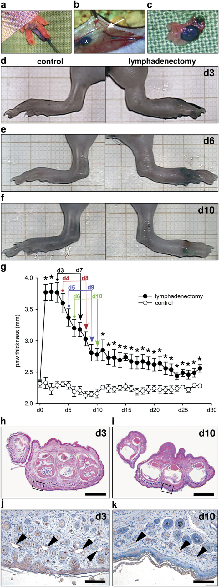
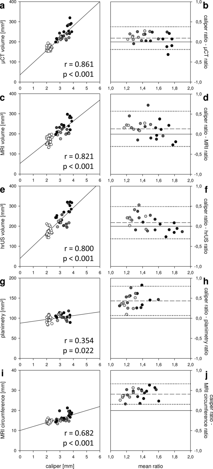
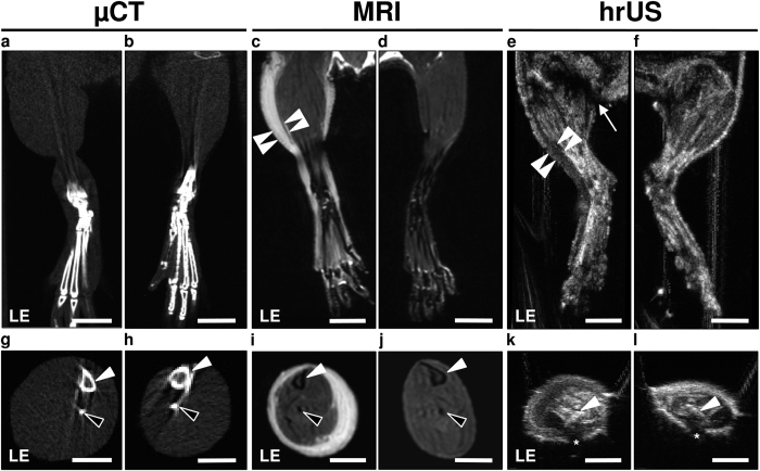
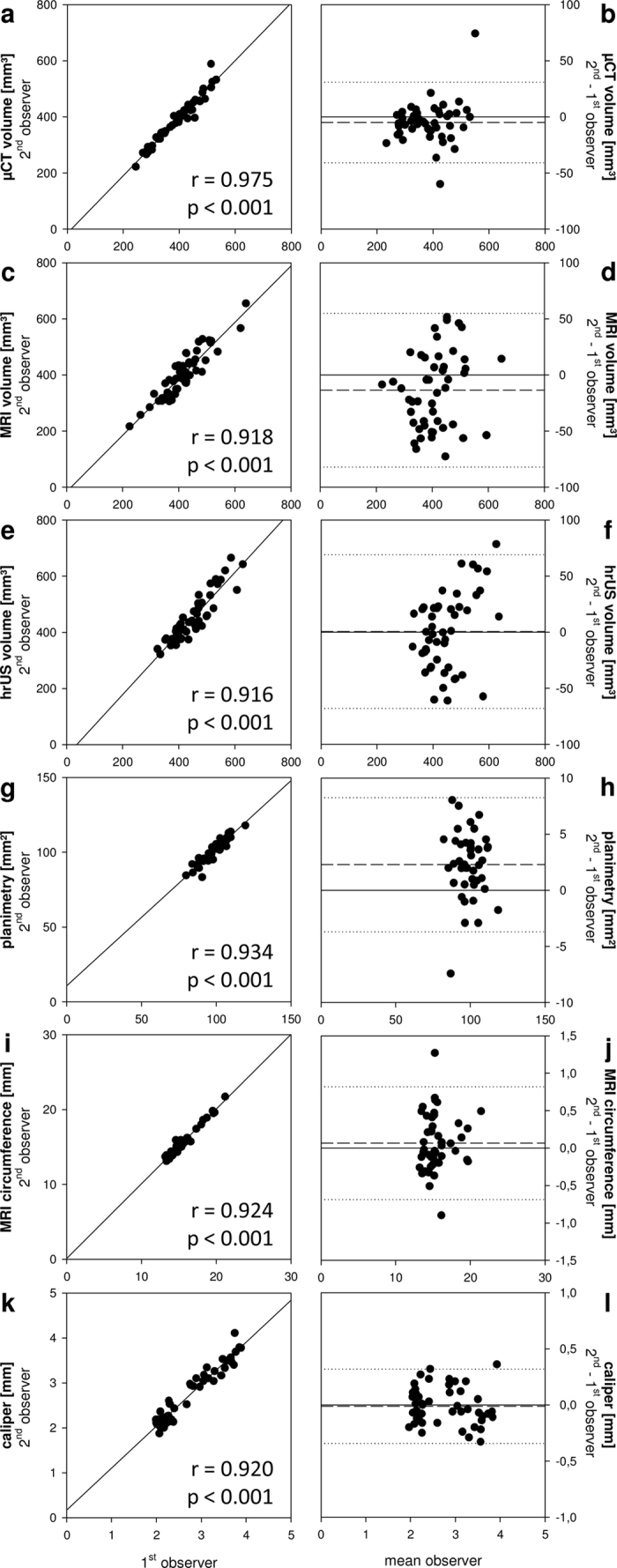
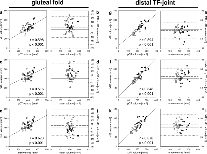
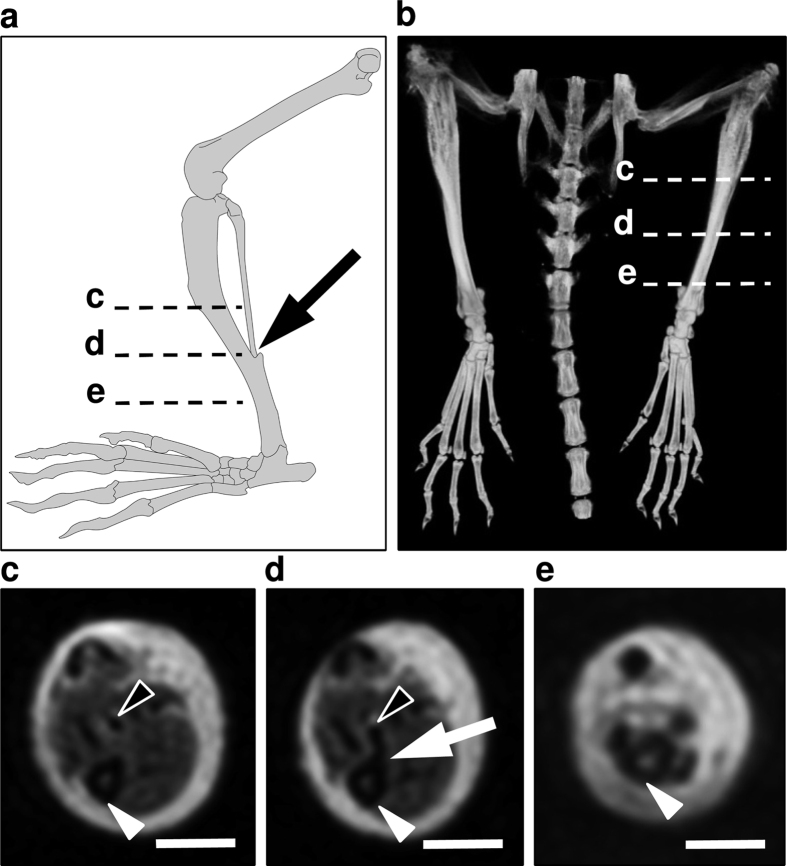
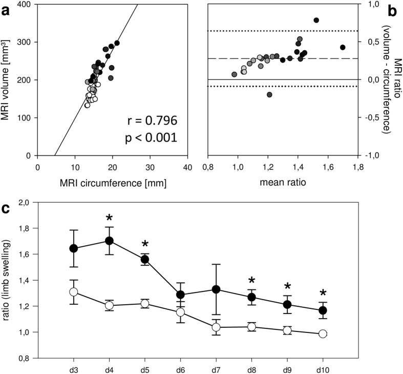
Secondary lymphedema is a common complication of cancer treatment characterized by chronic limb swelling with interstitial inflammation. The rodent hindlimb is a widely used model for the evaluation of novel lymphedema treatments. However, the assessment of limb volume in small animals is challenging. Recently, high-resolution three-dimensional (3D) imaging modalities have been introduced for rodent limb volumetry. In the present study we evaluated the validity of microcomputed tomography (μCT), magnetic resonance imaging (MRI) and ultrasound in comparison to conventional measuring techniques. For this purpose, acute lymphedema was induced in the mouse hindlimb by a modified popliteal lymphadenectomy. The 4-week course of this type of lymphedema was first assessed in 6 animals. In additional 12 animals, limb volumes were analyzed by μCT, 9.4 T MRI and 30 MHz ultrasound as well as by planimetry, circumferential length and paw thickness measurements. Interobserver correlation was high for all modalities, in particular for μCT analysis (r = 0.975, p < 0.001). Importantly, caliper-measured paw thickness correlated well with μCT (r = 0.861), MRI (r = 0.821) and ultrasound (r = 0.800). Because the assessment of paw thickness represents a time- and cost-effective approach, it may be ideally suited for the quantification of rodent hindlimb lymphedema.

摘要

继发性淋巴水肿是癌症治疗的常见并发症,其特征为慢性肢体肿胀伴间质炎症。啮齿动物后肢是评估新型淋巴水肿治疗方法的常用模型。然而,小动物肢体体积的评估具有挑战性。最近,高分辨率三维(3D)成像技术已被引入用于啮齿动物肢体体积测量。在本研究中,我们评估了微计算机断层扫描(μCT)、磁共振成像(MRI)和超声与传统测量技术相比的有效性。为此,通过改良的腘淋巴结切除术在小鼠后肢中诱导急性淋巴水肿。这种类型的淋巴水肿在 6 只动物中首先进行了 4 周的评估。在另外 12 只动物中,通过 μCT、9.4 T MRI 和 30 MHz 超声以及平面测量法、周径长度和爪厚度测量分析肢体体积。所有方法的观察者间相关性均较高,尤其是 μCT 分析(r=0.975,p<0.001)。重要的是,卡尺测量的爪厚度与 μCT(r=0.861)、MRI(r=0.821)和超声(r=0.800)相关性良好。由于爪厚度评估是一种省时且具有成本效益的方法,因此它可能非常适合量化啮齿动物后肢淋巴水肿。

https://cdn.ncbi.nlm.nih.gov/pmc/blobs/e1d0/5048170/08d44fd00c4b/srep34673-f7.jpg
https://cdn.ncbi.nlm.nih.gov/pmc/blobs/e1d0/5048170/93e146c0f653/srep34673-f1.jpg
https://cdn.ncbi.nlm.nih.gov/pmc/blobs/e1d0/5048170/e5209af0407f/srep34673-f2.jpg
https://cdn.ncbi.nlm.nih.gov/pmc/blobs/e1d0/5048170/610765112d53/srep34673-f3.jpg
https://cdn.ncbi.nlm.nih.gov/pmc/blobs/e1d0/5048170/0ae55a8244cd/srep34673-f4.jpg
https://cdn.ncbi.nlm.nih.gov/pmc/blobs/e1d0/5048170/b2e3ef88ddce/srep34673-f5.jpg
https://cdn.ncbi.nlm.nih.gov/pmc/blobs/e1d0/5048170/34be8b0b03c0/srep34673-f6.jpg
https://cdn.ncbi.nlm.nih.gov/pmc/blobs/e1d0/5048170/08d44fd00c4b/srep34673-f7.jpg

相似文献

1
High-resolution 3D volumetry versus conventional measuring techniques for the assessment of experimental lymphedema in the mouse hindlimb.高分辨率 3D 容积测量与传统测量技术在评估小鼠后肢实验性淋巴水肿中的比较。
Sci Rep. 2016 Oct 4;6:34673. doi: 10.1038/srep34673.
2
Microcomputed tomography versus plethysmometer and electronic caliper in the measurements of lymphedema in the hindlimb of mice.微计算机断层扫描与体积描记法和电子卡尺在测量小鼠后肢淋巴水肿中的比较。
Sci Rep. 2022 Jul 18;12(1):12267. doi: 10.1038/s41598-022-16311-2.
3
Magnetic Resonance Lymphography at 9.4 T Using a Gadolinium-Based Nanoparticle in Rats: Investigations in Healthy Animals and in a Hindlimb Lymphedema Model.9.4T 磁共振淋巴造影术在大鼠中使用基于钆的纳米颗粒:健康动物和后肢淋巴水肿模型的研究。
Invest Radiol. 2017 Dec;52(12):725-733. doi: 10.1097/RLI.0000000000000398.
4
Quantification of tissue volume in the hindlimb of mice using microcomputed tomography images and analysing software.使用 microCT 图像和分析软件对小鼠后肢组织体积进行定量分析。
Sci Rep. 2020 May 19;10(1):8297. doi: 10.1038/s41598-020-65214-7.
5
Radiation Inhibits Lymph Drainage in an Acquired Lymphedema Mouse Hindlimb Model.辐射抑制获得性淋巴水肿小鼠后肢模型中的淋巴引流。
Lymphat Res Biol. 2020 Feb;18(1):16-21. doi: 10.1089/lrb.2018.0072. Epub 2019 Jun 24.
6
Comparison between stromal vascular fraction and adipose derived stem cells in a mouse lymphedema model.基质血管成分与脂肪来源干细胞在小鼠淋巴水肿模型中的比较。
J Plast Surg Hand Surg. 2020 Oct;54(5):302-311. doi: 10.1080/2000656X.2020.1772799. Epub 2020 Jun 10.
7
Quantification of lymphedema in a rat model by 3D-active contour segmentation by magnetic resonance imaging.通过磁共振成像的三维活动轮廓分割对大鼠模型中的淋巴水肿进行定量分析。
Lymphat Res Biol. 2012 Mar;10(1):25-9. doi: 10.1089/lrb.2011.0010. Epub 2012 Mar 14.
8
Short term treatment of secondary lymphedema with hyaluronidase injections reduces mouse hindlimb lymphedema.透明质酸酶注射治疗继发性淋巴水肿的短期疗效可减轻小鼠后肢淋巴水肿。
J Plast Surg Hand Surg. 2023 Jun 20;58:40-47. doi: 10.2340/jphs.v58.7791.
9
Near-Infrared Fluorescence Imaging Directly Visualizes Lymphatic Drainage Pathways and Connections between Superficial and Deep Lymphatic Systems in the Mouse Hindlimb.近红外荧光成像直接可视化小鼠后肢浅深淋巴系统之间的淋巴引流途径和连接。
Sci Rep. 2018 May 4;8(1):7078. doi: 10.1038/s41598-018-25383-y.
10
An experimental model for chronic lymphedema.一种慢性淋巴水肿的实验模型。
Plast Reconstr Surg. 1990 Apr;85(4):573-80. doi: 10.1097/00006534-199004000-00012.

引用本文的文献

1
[Research progress on rodent models of secondary lymphedema].[继发性淋巴水肿啮齿动物模型的研究进展]
Zhongguo Xiu Fu Chong Jian Wai Ke Za Zhi. 2025 Jul 15;39(7):919-924. doi: 10.7507/1002-1892.202504094.
2
Mesenchymal stem cell therapy for breast cancer-related secondary lymphedema (Review).间充质干细胞治疗乳腺癌相关继发性淋巴水肿(综述)
Mol Clin Oncol. 2025 Jun 23;23(2):73. doi: 10.3892/mco.2025.2868. eCollection 2025 Aug.
3
Ultrasound guidance for intervertebral disc location and paravertebral tissue swelling quantification in a mouse model of intervertebral disc herniation.

本文引用的文献

1
Prevention of Postsurgical Lymphedema by 9-cis Retinoic Acid.9-顺式维甲酸预防术后淋巴水肿
Ann Surg. 2016 Aug;264(2):353-61. doi: 10.1097/SLA.0000000000001525.
2
Adipose-derived stem cell transplantation for therapeutic lymphangiogenesis in a mouse secondary lymphedema model.脂肪来源干细胞移植用于小鼠继发性淋巴水肿模型中的治疗性淋巴管生成
Regen Med. 2015;10(5):549-62. doi: 10.2217/rme.15.24.
3
Animal models in surgical lymphedema research--a systematic review.手术性淋巴水肿研究中的动物模型——一项系统评价。
在椎间盘突出症小鼠模型中,超声引导用于椎间盘定位及椎旁组织肿胀定量分析。
Eur Spine J. 2025 Apr 24. doi: 10.1007/s00586-025-08843-8.
4
3D scanner's potential as a novel tool for lymphedema measurement in mouse hindlimb models.3D扫描仪作为一种用于小鼠后肢模型淋巴水肿测量的新型工具的潜力。
Sci Rep. 2025 Jan 30;15(1):3747. doi: 10.1038/s41598-025-85637-4.
5
Methods of ex vivo analysis of tissue status in vascularized composite allografts.血管化复合异体移植物组织状态的离体分析方法。
J Transl Med. 2023 Sep 8;21(1):609. doi: 10.1186/s12967-023-04379-x.
6
Lymphatic channel sheet of polydimethylsiloxane for preventing secondary lymphedema in the rat upper limb model.用于预防大鼠上肢模型继发性淋巴水肿的聚二甲基硅氧烷淋巴通道片
Bioeng Transl Med. 2022 Jul 5;8(1):e10371. doi: 10.1002/btm2.10371. eCollection 2023 Jan.
7
Adipose tissue-derived microvascular fragments promote lymphangiogenesis in a murine lymphedema model.脂肪组织来源的微血管片段在小鼠淋巴水肿模型中促进淋巴管生成。
J Tissue Eng. 2022 Jul 26;13:20417314221109957. doi: 10.1177/20417314221109957. eCollection 2022 Jan-Dec.
8
Microcomputed tomography versus plethysmometer and electronic caliper in the measurements of lymphedema in the hindlimb of mice.微计算机断层扫描与体积描记法和电子卡尺在测量小鼠后肢淋巴水肿中的比较。
Sci Rep. 2022 Jul 18;12(1):12267. doi: 10.1038/s41598-022-16311-2.
9
Comparison of normal hindlimb lymphatic systems in rats with detours present after lymphatic flow blockage.比较淋巴管阻塞后出现旁路的大鼠正常后肢淋巴系统。
PLoS One. 2021 Dec 13;16(12):e0260404. doi: 10.1371/journal.pone.0260404. eCollection 2021.
10
Current Advancements in Animal Models of Postsurgical Lymphedema: A Systematic Review.术后淋巴水肿动物模型的最新进展:系统评价。
Adv Wound Care (New Rochelle). 2022 Aug;11(8):399-418. doi: 10.1089/wound.2021.0033. Epub 2021 Aug 27.
J Surg Res. 2016 Jan;200(1):208-20. doi: 10.1016/j.jss.2015.07.005. Epub 2015 Jul 9.
4
Microsurgical Lymphatic Vessel Transplantation.显微外科淋巴管移植
J Reconstr Microsurg. 2016 Jan;32(1):34-41. doi: 10.1055/s-0035-1554934. Epub 2015 Jul 10.
5
Quantity of lymph nodes correlates with improvement in lymphatic drainage in treatment of hind limb lymphedema with lymph node flap transfer in rats.在大鼠后肢淋巴水肿的淋巴结瓣转移治疗中,淋巴结数量与淋巴引流改善情况相关。
Microsurgery. 2016 Mar;36(3):239-45. doi: 10.1002/micr.22388. Epub 2015 Feb 25.
6
Prevention of Lymphedematous Change in the Mouse Hindlimb by Nonvascularized Lymph Node Transplantation.通过非血管化淋巴结移植预防小鼠后肢淋巴水肿性改变
Ann Plast Surg. 2016 Apr;76(4):442-5. doi: 10.1097/SAP.0000000000000428.
7
Bilateral lower extremity inflammatory lymphedema in Air Force basic trainees: clinical and epidemiologic study of a new disease entity.空军新兵中双侧下肢炎症性淋巴水肿:一种新疾病实体的临床和流行病学研究。
JAMA Dermatol. 2015 Apr;151(4):395-400. doi: 10.1001/jamadermatol.2014.3794.
8
Photoacoustic lymphatic imaging with high spatial-temporal resolution.具有高时空分辨率的光声淋巴成像
J Biomed Opt. 2014 Nov;19(11):116009. doi: 10.1117/1.JBO.19.11.116009.
9
Developing a Lower Limb Lymphedema Animal Model with Combined Lymphadenectomy and Low-dose Radiation.通过联合淋巴结切除术和低剂量辐射建立下肢淋巴水肿动物模型
Plast Reconstr Surg Glob Open. 2014 Apr 7;2(3):e121. doi: 10.1097/GOX.0000000000000064. eCollection 2014 Mar.
10
Sterile inflammation after lymph node transfer improves lymphatic function and regeneration.淋巴结移植后的无菌性炎症可改善淋巴管功能和再生。
Plast Reconstr Surg. 2014 Jul;134(1):60-68. doi: 10.1097/PRS.0000000000000286.