Foditsch Elena Esra, Zimmermann Reinhold
Urology, Spinal Cord Injury and Tissue Regeneration Center Salzburg, Paracelsus Medical University.
University Clinic of Urology and Andrology, Salzburg General Hospital, Paracelsus Medical University, Salzburg, Austria.
Res Rep Urol. 2016 Sep 28;8:169-173. doi: 10.2147/RRU.S115181. eCollection 2016.
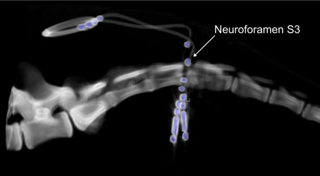
The aim of this study was to develop a controlled approach for sacral neuromodulation (SNM) to improve both nerve targeting and tined lead placement, for which a new computed tomography (CT)-guided implantation technique was analyzed in minipigs.
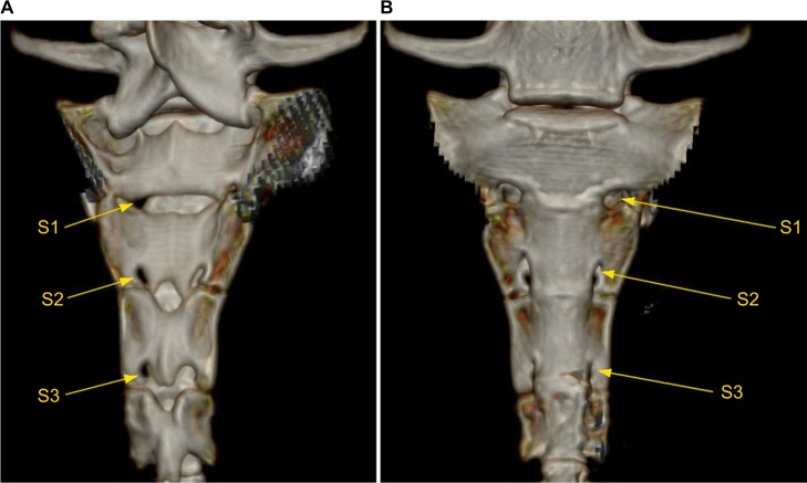
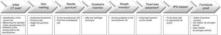
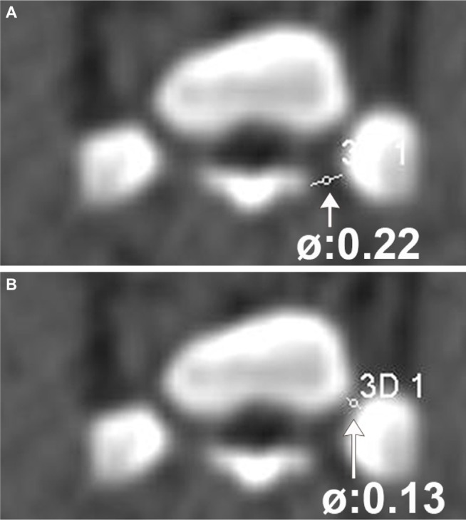
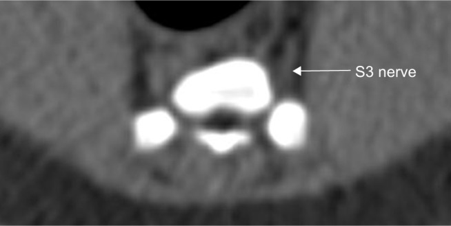
This study included five female, adult Göttingen minipigs. In deep sedoanalgesia, the minipigs were placed in an extended prone position. Commercially available SNM materials were used (needle, introduction sheath, and quadripolar tined lead electrode). Gross anatomy was displayed by CT, and the nerves were bilaterally identified. The optimal angles to puncture the S3 foramen, the resulting access path, and the site for the skin incision were defined subsequently. The needle puncture and the tined lead placement were followed by successive CT scans/3D-reconstruction images. Once proper CT-guided placement of the needle and electrode was established, response to functional stimuli was intraoperatively checked to verify correct positioning.
Successful bilateral tined lead implantation was performed in four out of five minipigs. Implantation was different from the clinical situation because the puncture was done from the contralateral side at a 30° angle to the midline and 60° horizontal angle to ensure both passage through the foramen and nerve access. Surgery time was 50-150 minutes. Stimulation response comprised a twitch of the perianal musculature and tail rotation to the contralateral side.
We have established a new, minimally invasive, highly standardized, CT-guided SNM electrode implantation technique. Functional outcomes are clearly defined and reproducible. All procedures can be performed without complications. Future chronic stimulation studies in minipigs can thereby be conducted using a controlled and highly standardized protocol.
本研究的目的是开发一种用于骶神经调节(SNM)的可控方法,以改善神经靶向和带倒刺导线的放置,为此在小型猪中分析了一种新的计算机断层扫描(CT)引导植入技术。
本研究包括5只成年雌性哥廷根小型猪。在深度镇静镇痛下,将小型猪置于伸展俯卧位。使用市售的SNM材料(针、导入鞘和四极带倒刺导线电极)。通过CT显示大体解剖结构,并双侧识别神经。随后确定穿刺S3孔的最佳角度、由此产生的进入路径以及皮肤切口的位置。在针穿刺和带倒刺导线放置后进行连续的CT扫描/三维重建图像。一旦通过CT引导确定针和电极放置正确,术中检查功能刺激的反应以验证定位是否正确。
5只小型猪中有4只成功进行了双侧带倒刺导线植入。植入与临床情况不同,因为穿刺是从对侧以与中线成30°角和水平成60°角进行的,以确保既通过孔又能接触神经。手术时间为50 - 150分钟。刺激反应包括肛周肌肉组织的抽搐和尾巴向对侧旋转。
我们建立了一种新的、微创的、高度标准化的CT引导SNM电极植入技术。功能结果明确且可重复。所有操作均无并发症。因此,未来可以使用可控且高度标准化的方案在小型猪中进行慢性刺激研究。