Nuvvula Sivakumar, Manepalli Swapna, Mohapatra Abinash, Mallineni Sreekanth Kumar
Department of Paedodontics and Preventive Dentistry, Narayana Dental College, Nellore, Andhra Pradesh 524003, India.
Case Rep Dent. 2016;2016:2319890. doi: 10.1155/2016/2319890. Epub 2016 Sep 22.
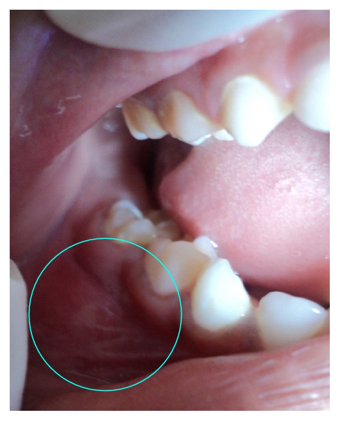
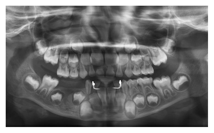
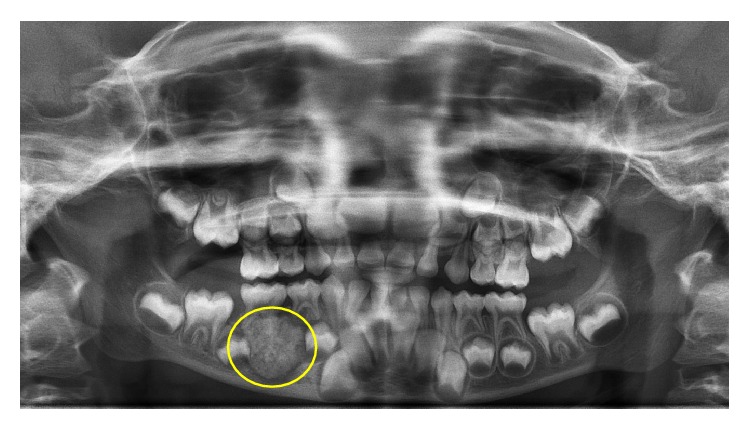
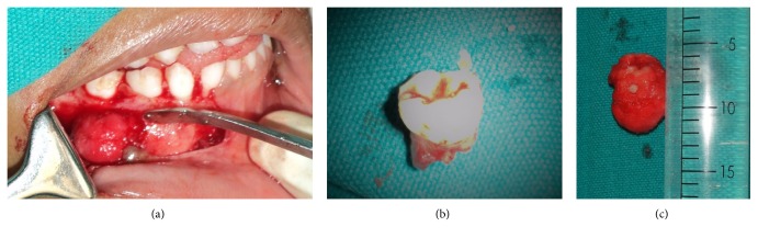
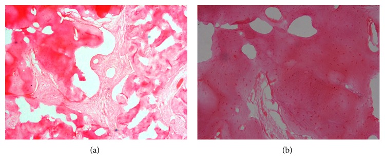
Cementoblastoma is a benign lesion of the odontogenic ectomesenchymal origin. It rarely occurs in primary dentition. This report describes a case of a cementoblastoma relating to the right mandibular second primary molar in a 7-year-old girl. Her panoramic radiograph revealed a well-defined radiopaque lesion with a radiolucent border extending from the distal surface of the mandibular right first primary molar to the distal surface of mandibular second primary molar. The tumor was attached to the mesial root of primary second molar and was excised along with the teeth involved and sent for histopathological evaluation, which showed irregular trabeculae of mineralized tissue interspersed with fibrovascular connective tissue, trabeculae of mineralized tissue with prominent reversal lines, and peripheral rimming of the mineralized tissue with blast cells. On a six-month follow-up, there has been no recurrence of the lesion.
成牙骨质细胞瘤是一种牙源性外胚间叶组织起源的良性病变。它很少发生于乳牙列。本报告描述了一例发生于一名7岁女孩右下颌第二乳磨牙的成牙骨质细胞瘤。她的全景片显示一个边界清晰的不透光病变,有一条透射边界,从右下颌第一乳磨牙远中面延伸至右下颌第二乳磨牙远中面。肿瘤附着于第二乳磨牙近中根,与受累牙齿一并切除并送去做组织病理学评估,结果显示矿化组织的不规则小梁间杂有纤维血管结缔组织、有明显反转线的矿化组织小梁,以及矿化组织周边有胚细胞环绕。在六个月的随访中,病变无复发。