Lim Beom Jin, Yang Jae Won, Do Woo Sung, Fogo Agnes B
Department of Pathology, Yonsei University College of Medicine, Seoul, Korea.
Department of Nephrology, Yonsei University Wonju College of Medicine, Wonju, Korea.
J Pathol Transl Med. 2016 Nov;50(6):405-410. doi: 10.4132/jptm.2016.09.21. Epub 2016 Oct 16.
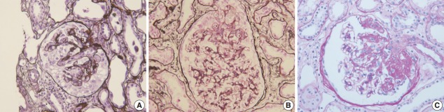
Focal segmental glomerulosclerosis (FSGS) is characterized by focal and segmental obliteration of glomerular capillary tufts with increased matrix. FSGS is classified as collapsing, tip, cellular, perihilar and not otherwise specified variants according to the location and character of the sclerotic lesion. Primary or idiopathic FSGS is considered to be related to podocyte injury, and the pathogenesis of podocyte injury has been actively investigated. Several circulating factors affecting podocyte permeability barrier have been proposed, but not proven to cause FSGS. FSGS may also be caused by genetic alterations. These genes are mainly those regulating slit diaphragm structure, actin cytoskeleton of podocytes, and foot process structure. The mode of inheritance and age of onset are different according to the gene involved. Recently, the role of parietal epithelial cells (PECs) has been highlighted. Podocytes and PECs have common mesenchymal progenitors, therefore, PECs could be a source of podocyte repopulation after podocyte injury. Activated PECs migrate along adhesion to the glomerular tuft and may also contribute to the progression of sclerosis. Markers of activated PECs, including CD44, could be used to distinguish FSGS from minimal change disease. The pathogenesis of FSGS is very complex; however, understanding basic mechanisms of podocyte injury is important not only for basic research, but also for daily diagnostic pathology practice.
局灶节段性肾小球硬化(FSGS)的特征是肾小球毛细血管袢局灶性和节段性闭塞伴基质增加。根据硬化病变的部位和特征,FSGS可分为塌陷型、顶端型、细胞型、肾小球旁型及未分类型。原发性或特发性FSGS被认为与足细胞损伤有关,且对足细胞损伤的发病机制已进行了积极研究。已提出几种影响足细胞通透屏障的循环因子,但尚未证实其可导致FSGS。FSGS也可能由基因改变引起。这些基因主要是调节裂孔隔膜结构、足细胞肌动蛋白细胞骨架和足突结构的基因。根据所涉及的基因不同,遗传方式和发病年龄也有所不同。最近,壁层上皮细胞(PEC)的作用受到了关注。足细胞和PEC有共同的间充质祖细胞,因此,PEC可能是足细胞损伤后足细胞再填充的来源。活化的PEC沿黏附迁移至肾小球袢,也可能促进硬化的进展。活化PEC的标志物,包括CD44,可用于鉴别FSGS和微小病变肾病。FSGS的发病机制非常复杂;然而,了解足细胞损伤的基本机制不仅对基础研究很重要,对日常诊断病理实践也很重要。