Shimura Yuta, Sato Yuji, Kitagawa Noboru, Omori Miyuki
Department of Geriatric Dentistry, Showa University, 2-1-1 Kitasenzoku, Ota-ku, Tokyo, 145-8515, Japan.
Int J Implant Dent. 2016 Dec;2(1):17. doi: 10.1186/s40729-016-0050-6. Epub 2016 Jun 17.
Proper implant placement is very important for long-term implant stability. Recently, numerous biomechanical studies have been conducted to clarify the relationship between implant placement and peri-implant stress. The placement of multiple implants in the edentulous posterior mandible has been studied by geometric analysis, three-dimensional finite element analysis (FEA), model experimentation, etc. Offset placement is a technique that reduces peri-implant load. However, few studies have used multiple analyses to clarify the value of the offset placement under identical conditions. The present study aimed to clarify the biomechanical effects of offset placement on the peri-implant bone in edentulous posterior mandibles by comparative investigation using FEA and model experimentation with strain gauges.
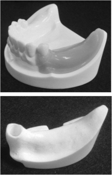
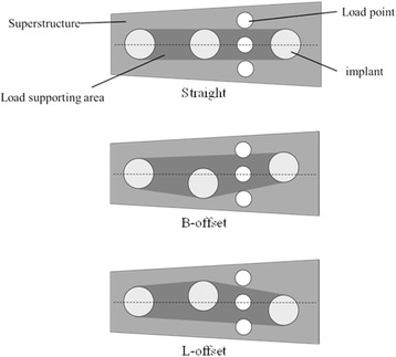
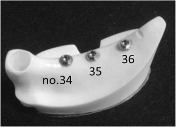
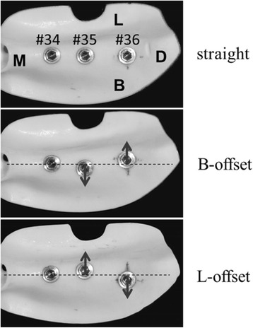
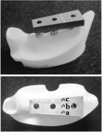
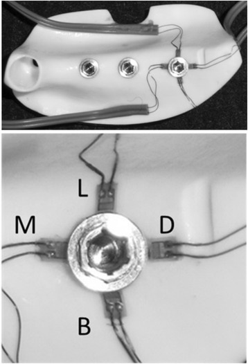
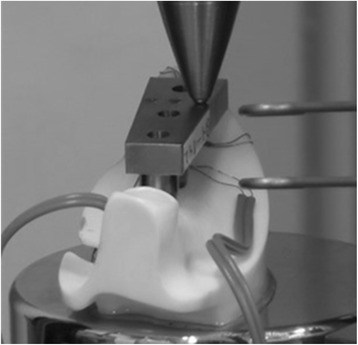
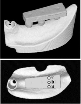
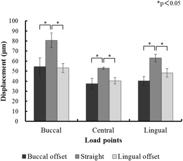
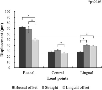
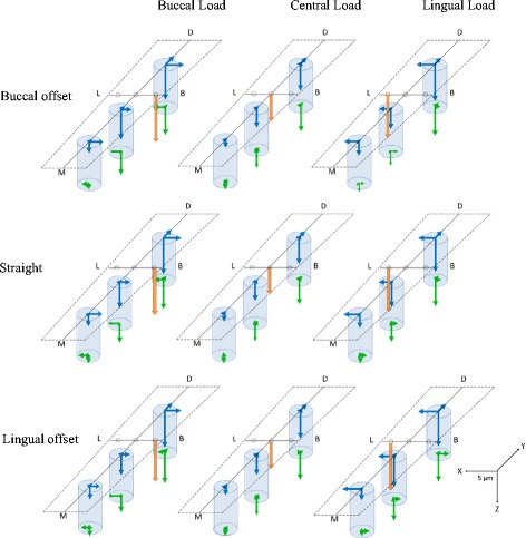
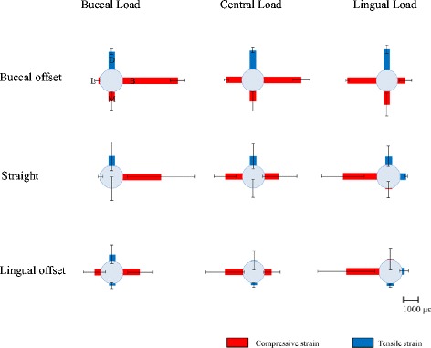
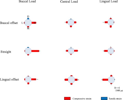
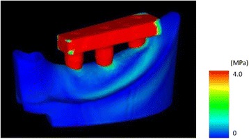
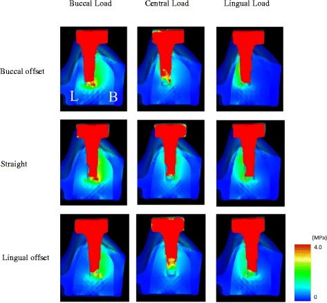
Three implants were embedded in an artificial mandible in the parts corresponding to the first premolar, the second premolar, and the first molar. A titanium superstructure was mounted to prepare models (experimental models). Three load points (buccal, central, and lingual) were established on the part of the superstructure corresponding to the first molar. Three types of experimental models, each with a different implant placement, were prepared. In one model, the implants were placed in a straight line; in the other two, the implants in the parts corresponding to the second premolar and the first molar were offset each by a 1-mm increment to the buccal or lingual side. Four strain gauges were applied to the peri-implant bone corresponding to the first molar. The experimental models were imaged by micro-computed tomography (CT), and FEA models were constructed from the CT data. A vertical load of 100 N was applied on the three load points in the experimental models and in the FEA models. The extent of compressed displacement and the strain in the peri-implant bone were compared between the experimental models and the FEA models.
Both experimental and FEA models suffered the least compressed displacement during central loading in all placements. The greatest stress and compressive strain was on the load side in all types of placements.
Offset placement may not necessarily be more biomechanically effective than straight placement in edentulous posterior mandibles.
正确的种植体植入对于种植体的长期稳定性非常重要。最近,已经进行了大量的生物力学研究来阐明种植体植入与种植体周围应力之间的关系。通过几何分析、三维有限元分析(FEA)、模型实验等方法对无牙后下颌骨中多个种植体的植入进行了研究。偏移植入是一种减少种植体周围负荷的技术。然而,很少有研究使用多种分析方法来阐明在相同条件下偏移植入的价值。本研究旨在通过使用FEA和带应变片的模型实验进行对比研究,阐明偏移植入对无牙后下颌骨种植体周围骨的生物力学影响。
在人工下颌骨中对应于第一前磨牙、第二前磨牙和第一磨牙的部位植入三颗种植体。安装钛制上部结构以制备模型(实验模型)。在对应于第一磨牙的上部结构部分设置三个加载点(颊侧、中央和舌侧)。制备三种不同种植体植入方式的实验模型。在一个模型中,种植体呈直线排列;在另外两个模型中,对应于第二前磨牙和第一磨牙部位的种植体分别向颊侧或舌侧偏移1毫米。在对应于第一磨牙的种植体周围骨上应用四个应变片。通过微计算机断层扫描(CT)对实验模型进行成像,并根据CT数据构建FEA模型。在实验模型和FEA模型的三个加载点上施加100 N的垂直载荷。比较实验模型和FEA模型中种植体周围骨的压缩位移程度和应变。
在所有植入方式中,实验模型和FEA模型在中央加载时压缩位移最小。在所有类型的植入方式中,最大应力和压缩应变都出现在加载侧。
在无牙后下颌骨中,偏移植入在生物力学方面不一定比直线植入更有效。