Ford Neil C, Baccei Mark L
Neuroscience Graduate Program, University of Cincinnati College of Medicine, 231 Albert Sabin Way, Cincinnati, OH 45267, USA; Pain Research Center, Department of Anesthesiology, University of Cincinnati Medical Center, 231 Albert Sabin Way, Cincinnati, OH 45267, USA.
Neuroscience Graduate Program, University of Cincinnati College of Medicine, 231 Albert Sabin Way, Cincinnati, OH 45267, USA; Pain Research Center, Department of Anesthesiology, University of Cincinnati Medical Center, 231 Albert Sabin Way, Cincinnati, OH 45267, USA.
Neuroscience. 2016 Dec 17;339:502-510. doi: 10.1016/j.neuroscience.2016.10.027. Epub 2016 Oct 14.
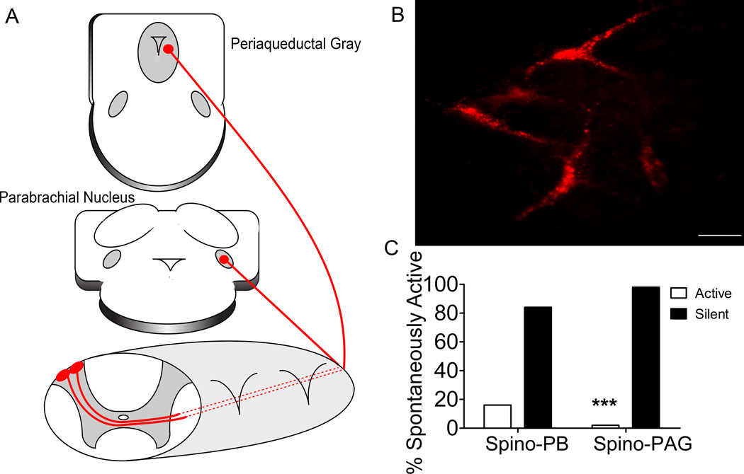
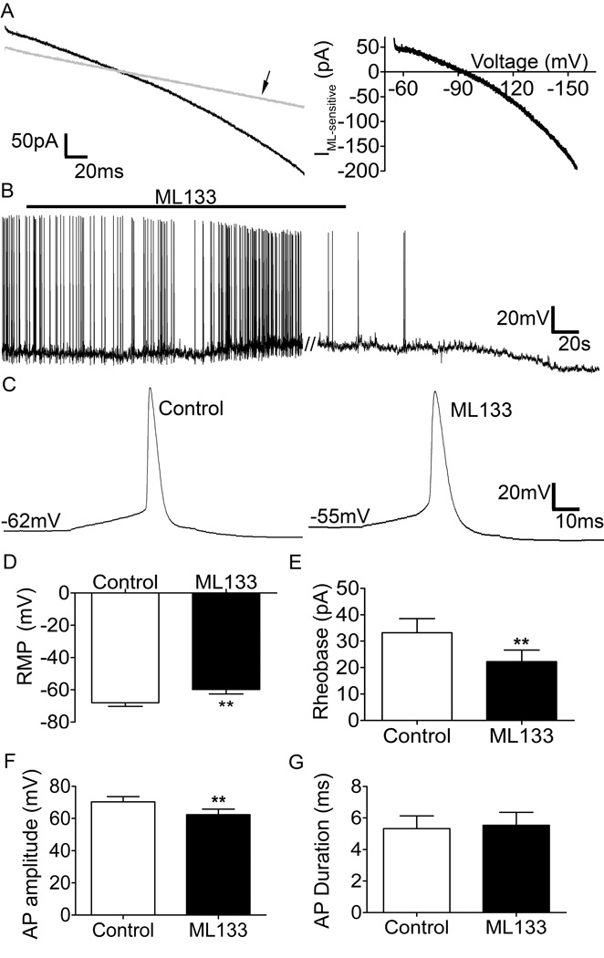
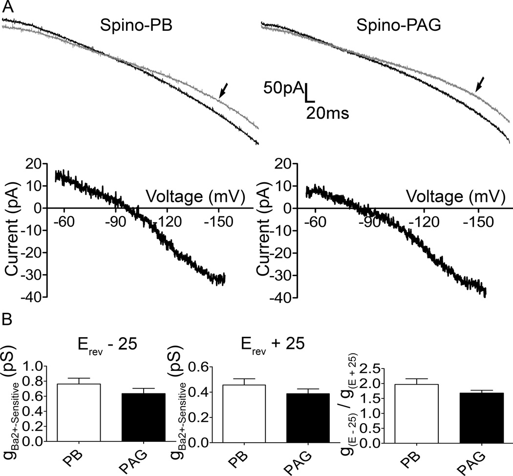
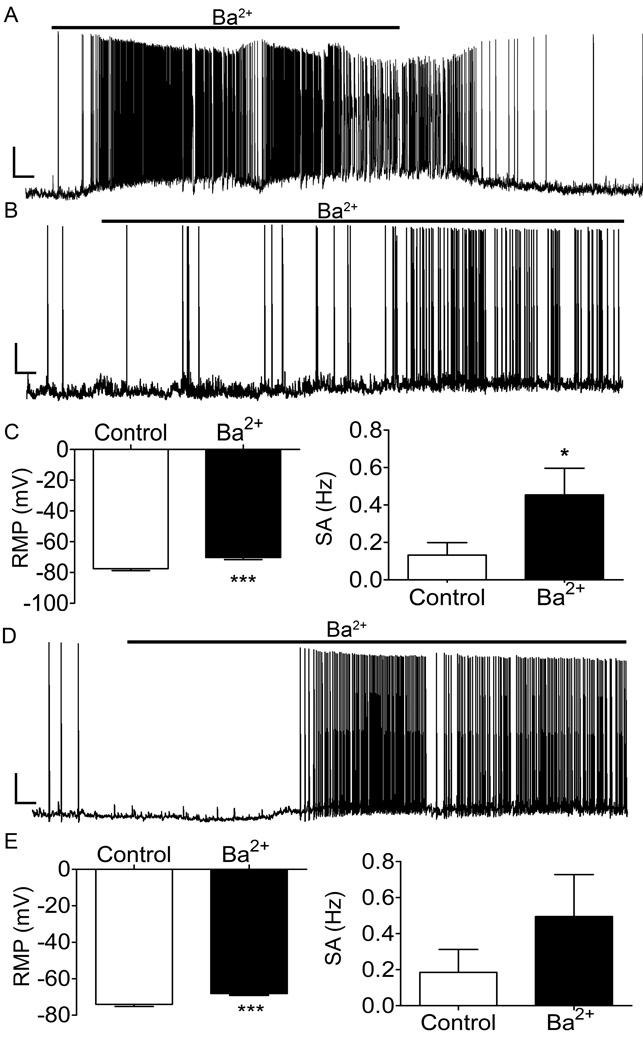
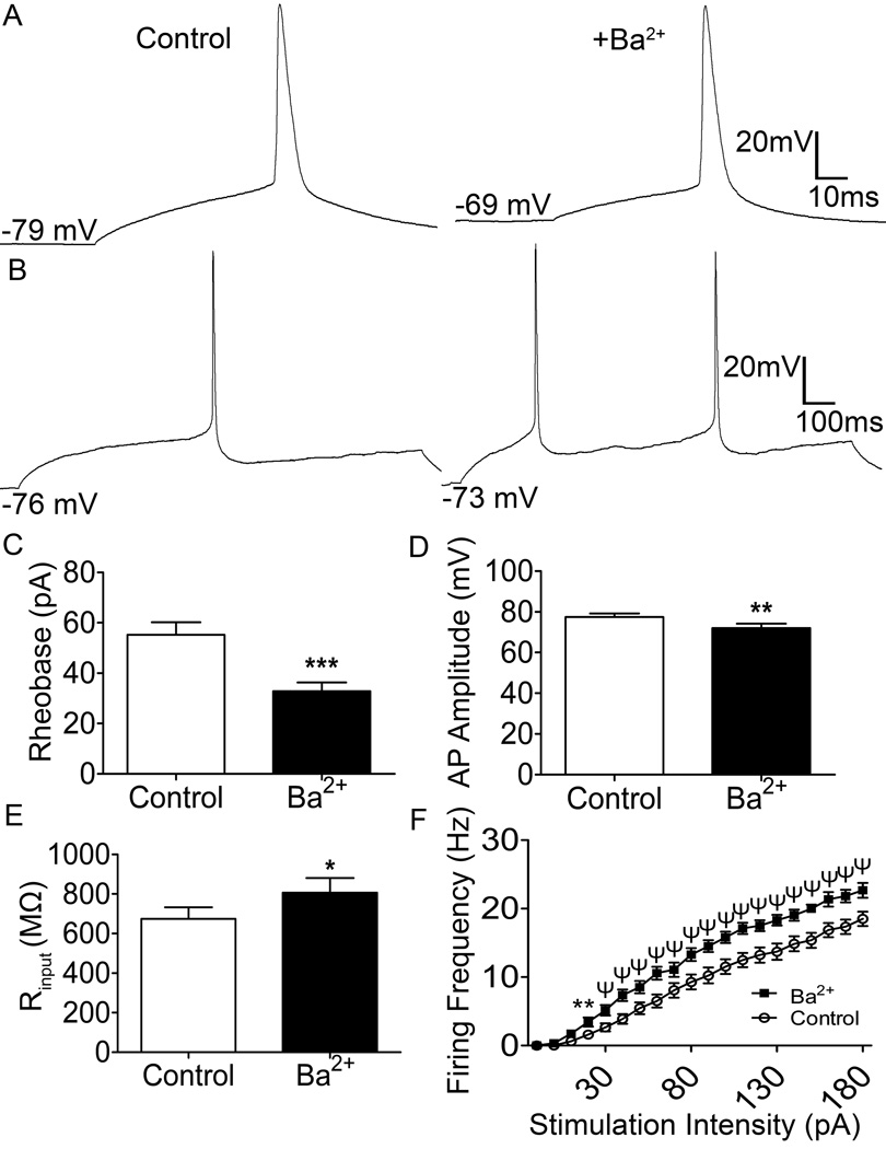
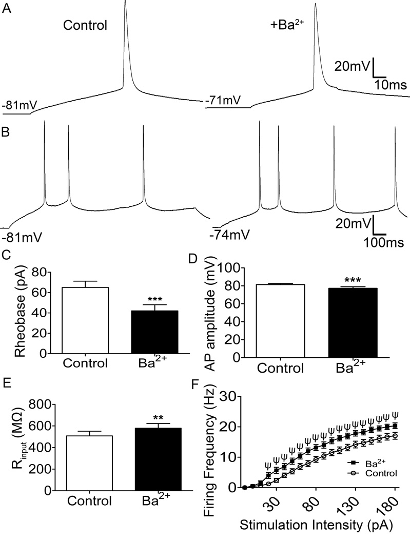
Spinal lamina I projection neurons serve as a major conduit by which noxious stimuli detected in the periphery are transmitted to nociceptive circuits in the brain, including the parabrachial nucleus (PB) and the periaqueductal gray (PAG). While neonatal spino-PB neurons are more than twice as likely to exhibit spontaneous activity compared to spino-PAG neurons, the underlying mechanisms remain unclear since nothing is known about the voltage-independent (i.e. 'leak') ion channels expressed by these distinct populations during early life. To begin identifying these key leak conductances, the present study investigated the role of classical inward-rectifying K (K2) channels in the regulation of intrinsic excitability in neonatal rat spino-PB and spino-PAG neurons. The data demonstrate that a reduction in K2-mediated conductance by external BaCl significantly enhanced intrinsic membrane excitability in both groups. Similar results were observed in spino-PB neurons following K2 channel block with the selective antagonist ML133. In addition, voltage-clamp experiments showed that spino-PB and spino-PAG neurons express similar amounts of K2 current during the early postnatal period, suggesting that the differences in the prevalence of spontaneous activity between the two populations are not explained by differential expression of K2 channels. Overall, the results indicate that K2-mediated conductance tonically dampens the firing of multiple subpopulations of lamina I projection neurons during early life. Therefore, K2 channels are positioned to tightly shape the output of the immature spinal nociceptive circuit and thus regulate the ascending flow of nociceptive information to the developing brain, which has important functional implications for pediatric pain.
脊髓板层I投射神经元是外周检测到的伤害性刺激传递至大脑伤害性感受回路(包括臂旁核(PB)和导水管周围灰质(PAG))的主要通道。虽然新生期脊髓-臂旁核神经元表现出自发活动的可能性是脊髓-导水管周围灰质神经元的两倍多,但潜在机制仍不清楚,因为对于这些不同神经元群体在生命早期所表达的电压非依赖性(即“泄漏”)离子通道一无所知。为了开始确定这些关键的泄漏电导,本研究调查了经典内向整流钾(K2)通道在调节新生大鼠脊髓-臂旁核和脊髓-导水管周围灰质神经元内在兴奋性中的作用。数据表明,通过外部氯化钡降低K2介导的电导可显著增强两组神经元的内在膜兴奋性。在用选择性拮抗剂ML133阻断K2通道后,在脊髓-臂旁核神经元中也观察到了类似结果。此外,电压钳实验表明,在出生后早期,脊髓-臂旁核和脊髓-导水管周围灰质神经元表达的K2电流数量相似,这表明这两个神经元群体之间自发活动发生率的差异不能用K2通道的差异表达来解释。总体而言,结果表明K2介导的电导在生命早期对板层I投射神经元多个亚群的放电起紧张性抑制作用。因此,K2通道对未成熟脊髓伤害性感受回路的输出进行严格塑形,从而调节伤害性信息向发育中大脑的上行传递,这对小儿疼痛具有重要的功能意义。