Albright Benjamin, Dhaher Roni, Wang Helen, Harb Roa, Lee Tih-Shih W, Zaveri Hitten, Eid Tore
Department of Laboratory Medicine, Yale University School of Medicine, New Haven, CT 06520, USA.
Department of Psychiatry, Yale University School of Medicine, New Haven, CT 06520, USA.
Exp Neurol. 2017 Feb;288:122-133. doi: 10.1016/j.expneurol.2016.10.007. Epub 2016 Oct 18.
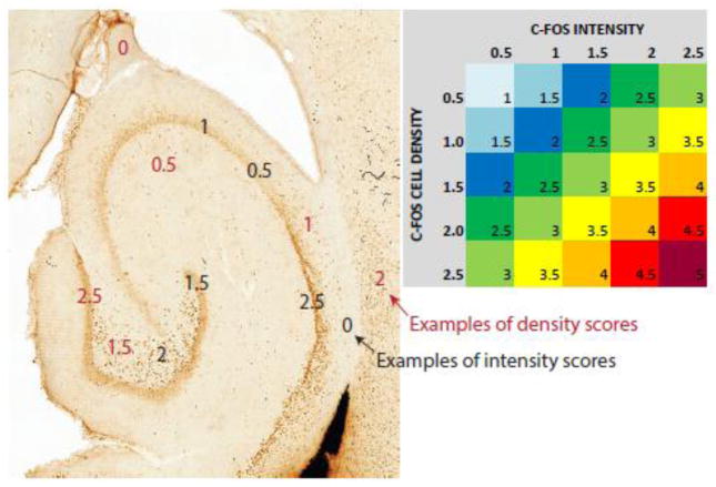
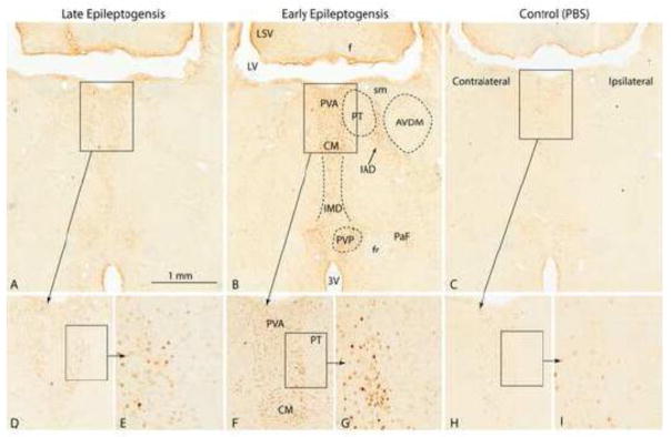
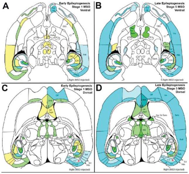
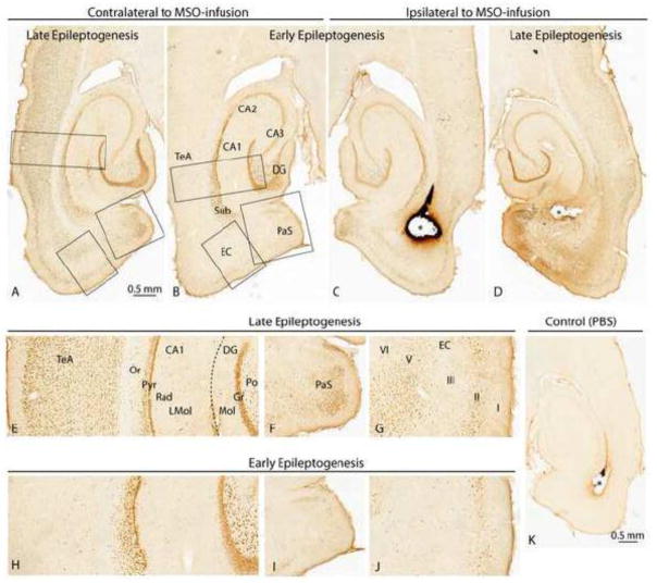
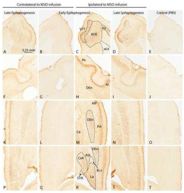
Loss of glutamine synthetase (GS) in hippocampal astrocytes has been implicated in the causation of human mesial temporal lobe epilepsy (MTLE). However, the mechanism by which the deficiency in GS leads to epilepsy is incompletely understood. Here we ask how hippocampal GS inhibition affects seizure phenotype and neuronal activation during epilepsy development (epileptogenesis). Epileptogenesis was induced by infusing the irreversible GS blocker methionine sulfoximine (MSO) unilaterally into the hippocampal formation of rats. We then used continuous video-intracranial electroencephalogram (EEG) monitoring and c-Fos immunohistochemistry to determine the type of seizures and spatial distribution of neuronal activation early (1-5days postinfusion) and late (16-43days postinfusion) in epileptogenesis. Early in epileptogenesis, seizures were preferentially mild (stage 1-2), activating neurons in the entorhinal-hippocampal area, the basolateral amygdala, the piriform cortex, the midline thalamus, and the anterior olfactory area. Late in epileptogenesis, the seizures were generally more severe (stages 4-5) with neuronal activation extending to the neocortex, the bed nucleus of the stria terminalis, the mediodorsal thalamu\s, and the central nucleus of the amygdala. Our findings demonstrate that inhibition of GS focally in the hippocampal formation triggers a process of epileptogenesis characterized by gradual worsening of seizure severity and involvement of progressively larger neuronal populations over a period of several weeks. Knowledge about the underlying mechanism of epileptogenesis is important because such knowledge may result in more specific and efficacious treatments of MTLE by moving away from large and poorly specific surgical resections to highly targeted surgical or pharmacological interventions of the epileptogenic process.
海马星形胶质细胞中谷氨酰胺合成酶(GS)的缺失与人类内侧颞叶癫痫(MTLE)的病因有关。然而,GS缺乏导致癫痫的机制尚未完全明确。在此,我们探讨海马GS抑制如何影响癫痫发展(癫痫发生)过程中的癫痫发作表型和神经元激活。通过向大鼠海马结构单侧注入不可逆的GS阻断剂蛋氨酸亚砜胺(MSO)诱导癫痫发生。然后,我们使用连续视频颅内脑电图(EEG)监测和c-Fos免疫组织化学来确定癫痫发生早期(注入后1-5天)和晚期(注入后16-43天)癫痫发作的类型以及神经元激活的空间分布。在癫痫发生早期,癫痫发作多为轻度(1-2期),激活内嗅-海马区、基底外侧杏仁核、梨状皮层、中线丘脑和前嗅区的神经元。在癫痫发生晚期,癫痫发作通常更严重(4-5期),神经元激活扩展到新皮层、终纹床核、丘脑背内侧核和杏仁核中央核。我们的研究结果表明,海马结构局部GS抑制引发了一个癫痫发生过程,其特征是癫痫发作严重程度逐渐恶化,且在数周内涉及的神经元群体逐渐增多。了解癫痫发生的潜在机制很重要,因为此类知识可能会使MTLE的治疗从大型且特异性差的手术切除转向对癫痫发生过程进行高度靶向的手术或药物干预,从而实现更具特异性和有效性的治疗。