Sun Xiaodong, Yan Wei, Zhang Jing, Niu Xiaoqing, Li Fuheng, Qin Weiquan, Ma Guangchang
Hainan Key Laboratory of Tropical Oil Crops Biology/Coconuts Research Institute, Chinese Academy of Tropical Agricultural Sciences, Wenchang, 571339 Hainan People's Republic of China ; College of Science, Northeast Agricultural University, Harbin, 150030 Heilongjiang Province People's Republic of China.
Hainan Key Laboratory of Tropical Oil Crops Biology/Coconuts Research Institute, Chinese Academy of Tropical Agricultural Sciences, Wenchang, 571339 Hainan People's Republic of China.
Springerplus. 2016 Oct 7;5(1):1748. doi: 10.1186/s40064-016-3416-6. eCollection 2016.
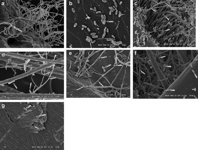
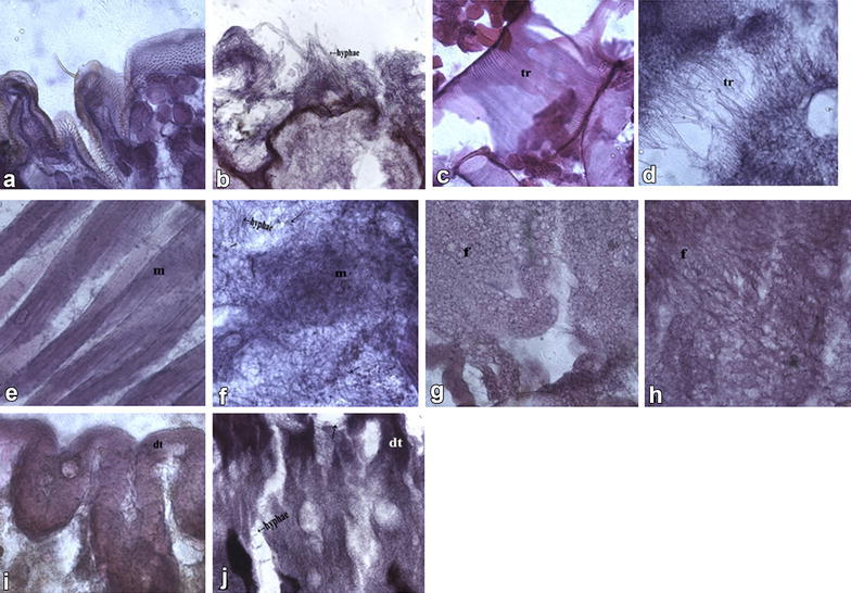
This study determined the pathogenicity of strain SD-3 against invasive red palm weevil (RPW), Olivier (coleoptera:curculionidae) larvae in Hainan Province, China. Inoculation of 1 × 10 conidia/mL caused 100 % mortality of , indicating that the conidia of strain SD-3 were highly virulent. The process of invasion mechanism was showed by scanning electron microscopy (SEM) and frozen section as follows. Once was infected by strain SD-3, hyphae first invaded the cuticular and body cavity of . Secondly, well-developed muscles, fat, tracheaes and digestive tube tissues in the abdomen of were then decomposed and absorbed by hyphae, leading to the total destruction of the larvae. Finally, hyphae reproduced, resulting in a large number of conidia in the body of RPW. The SEM and frozen section are convenient tools to observe the mode of action of entomopathogenic fungi and to observe how is able to colonize and infect the host.
本研究测定了菌株SD - 3对中国海南省红棕象甲幼虫(鞘翅目:象甲科)的致病性。接种1×10分生孢子/毫升导致红棕象甲幼虫100%死亡,表明菌株SD - 3的分生孢子具有高毒力。通过扫描电子显微镜(SEM)和冰冻切片显示的侵染机制过程如下。红棕象甲幼虫一旦被菌株SD - 3感染,菌丝首先侵入其表皮和体腔。其次,红棕象甲幼虫腹部发育良好的肌肉、脂肪、气管和消化道组织随后被菌丝分解和吸收,导致幼虫完全被破坏。最后,菌丝繁殖,导致红棕象甲体内产生大量分生孢子。扫描电子显微镜和冰冻切片是观察昆虫病原真菌作用方式以及观察其如何定殖和感染宿主的便捷工具。