Alshikho Mohamad J, Zürcher Nicole R, Loggia Marco L, Cernasov Paul, Chonde Daniel B, Izquierdo Garcia David, Yasek Julia E, Akeju Oluwaseun, Catana Ciprian, Rosen Bruce R, Cudkowicz Merit E, Hooker Jacob M, Atassi Nazem
From A.A. Martinos Center for Biomedical Imaging, Department of Radiology (M.J.A., N.R.Z., M.L.L., D.B.C., D.I.G., C.C., B.R.R., J.M.H.), Neurological Clinical Research Institute, Department of Neurology (M.J.A., P.C., J.E.Y., M.E.C., N.A.), and Department of Anesthesiology (O.A.), Massachusetts General Hospital, Harvard Medical School, Charlestown.
Neurology. 2016 Dec 13;87(24):2554-2561. doi: 10.1212/WNL.0000000000003427. Epub 2016 Nov 11.
In this cross-sectional study, we aimed to evaluate brain structural abnormalities in relation to glial activation in the same cohort of participants.
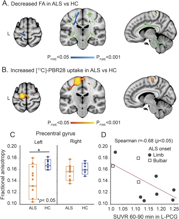
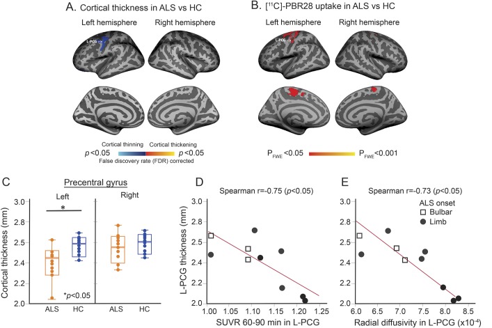
Ten individuals with amyotrophic lateral sclerosis (ALS) and 10 matched healthy controls underwent brain imaging using integrated MR/PET and the radioligand [C]-PBR28. Diagnosis history and clinical assessments including Upper Motor Neuron Burden Scale (UMNB) were obtained from patients with ALS. Diffusion tensor imaging (DTI) analyses including tract-based spatial statistics and tractography were applied. DTI metrics including fractional anisotropy (FA) and diffusivities (mean, axial, and radial) were measured in regions of interest. Cortical thickness was assessed using surface-based analysis. The locations of structural changes, measured by DTI and the areas of cortical thinning, were compared to regional glial activation measured by relative [C]-PBR28 uptake.
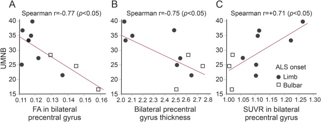
In this cohort of individuals with ALS, reduced FA and cortical thinning colocalized with regions demonstrating higher radioligand binding. [C]-PBR28 binding in the left motor cortex was correlated with FA (r = -0.68, p < 0.05) and cortical thickness (r = -0.75, p < 0.05). UMNB was correlated with glial activation (r = +0.75, p < 0.05), FA (r = -0.77, p < 0.05), and cortical thickness (r = -0.75, p < 0.05) in the motor cortex.
Increased uptake of the glial marker [C]-PBR28 colocalizes with changes in FA and cortical thinning. This suggests a link between disease mechanisms (gliosis and inflammation) and structural changes (cortical thinning and white and gray matter changes). In this multimodal neuroimaging work, we provide an in vivo model to investigate the pathogenesis of ALS.
在这项横断面研究中,我们旨在评估同一组参与者中与胶质细胞激活相关的脑结构异常。
10名肌萎缩侧索硬化症(ALS)患者和10名匹配的健康对照者接受了使用集成MR/PET和放射性配体[C]-PBR28的脑成像检查。从ALS患者处获取诊断病史和包括上运动神经元负荷量表(UMNB)在内的临床评估。应用了包括基于束的空间统计学和纤维束成像的扩散张量成像(DTI)分析。在感兴趣区域测量了包括分数各向异性(FA)和扩散率(平均、轴向和径向)在内的DTI指标。使用基于表面的分析评估皮质厚度。将通过DTI测量的结构变化位置和皮质变薄区域与通过相对[C]-PBR28摄取测量的区域胶质细胞激活进行比较。
在这组ALS患者中,FA降低和皮质变薄与显示较高放射性配体结合的区域共定位。左运动皮质中的[C]-PBR28结合与FA(r = -0.68,p < 0.05)和皮质厚度(r = -0.75,p < 0.05)相关。UMNB与运动皮质中的胶质细胞激活(r = +0.75,p < 0.05)、FA(r = -0.77,p < 0.05)和皮质厚度(r = -0.75,p < 0.05)相关。
胶质细胞标志物[C]-PBR28摄取增加与FA变化和皮质变薄共定位。这表明疾病机制(胶质细胞增生和炎症)与结构变化(皮质变薄以及白质和灰质变化)之间存在联系。在这项多模态神经影像学研究中,我们提供了一个体内模型来研究ALS的发病机制。