Fuglset Tone Seim, Endestad Tor, Hilland Eva, Bang Lasse, Tamnes Christian Krog, Landrø Nils Inge, Rø Øyvind
Regional Department for Eating Disorders, Division of Mental Health and Addiction, Oslo University Hospital, Ullevål, Oslo, Norway.
Department of Psychology, University of Oslo, Oslo, Norway.
BMC Psychiatry. 2016 Nov 16;16(1):404. doi: 10.1186/s12888-016-1126-9.
Anorexia nervosa (AN) is a severe mental illness, with an unknown etiology. Magnetic resonance imaging studies show reduced brain volumes and cortical thickness in patients compared to healthy controls. However, findings are inconsistent, especially concerning the anatomical location and extent of the differences. The purpose of this study was to estimate and compare brain volumes and regional cortical thickness in young females with AN and healthy controls.
Magnetic resonance imaging data was acquired from young females with anorexia nervosa (n = 23) and healthy controls (n = 28). Two different scanner sites were used. BMI varied from 13.5 to 20.7 within the patient group, and 11 patients had a BMI > 17.5. FreeSurfer was used to estimate brain volumes and regional cortical thickness.
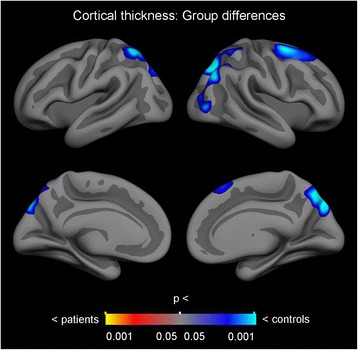
There were no differences between groups in total cerebral cortex volume, white matter volume, or lateral ventricle volume. There were also no volume differences in subcortical grey matter structures. However the results showed reduced cortical thickness bilaterally in the superior parietal gyrus, and in the right inferior parietal and superior frontal gyri.
The functional significance of the findings is undetermined as the majority of the included patients was already partially weight-restored. We discuss whether these regions could be related to predisposing factors of the illness, or whether they are regions that are more vulnerable to starvation, malnutrition or associated processes in AN.
神经性厌食症(AN)是一种严重的精神疾病,病因不明。磁共振成像研究表明,与健康对照相比,患者的脑容量和皮质厚度减小。然而,研究结果并不一致,尤其是在差异的解剖位置和范围方面。本研究的目的是评估和比较患有神经性厌食症的年轻女性与健康对照的脑容量和区域皮质厚度。
从患有神经性厌食症的年轻女性(n = 23)和健康对照(n = 28)获取磁共振成像数据。使用了两个不同的扫描部位。患者组的BMI在13.5至20.7之间变化,11名患者的BMI> 17.5。使用FreeSurfer来评估脑容量和区域皮质厚度。
两组在总大脑皮质体积、白质体积或侧脑室体积方面没有差异。皮质下灰质结构的体积也没有差异。然而,结果显示双侧顶上叶、右侧顶下叶和额上回的皮质厚度减小。
由于大多数纳入的患者已经部分恢复体重,这些发现的功能意义尚未确定。我们讨论了这些区域是否可能与该疾病的易感因素有关,或者它们是否是在神经性厌食症中更容易受到饥饿、营养不良或相关过程影响的区域。