Am J Psychiatry. 2013 Oct;170(10):1152-60. doi: 10.1176/appi.ajp.2013.12101294.
The pathophysiology of anorexia nervosa remains obscure, but structural brain alterations could be functionally important biomarkers. The authors assessed taste pleasantness and reward sensitivity in relation to brain structure, which may be related to food avoidance commonly seen in eating disorders.
The authors used structural MR imaging to study gray and white matter volumes in women with current restricting-type anorexia nervosa (N=19), women recovered from restricting-type anorexia nervosa (N=24), women with bulimia nervosa (N=19), and healthy comparison women (N=24).
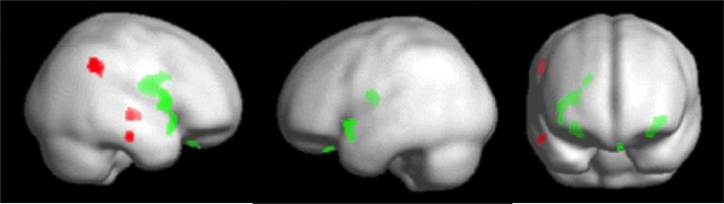
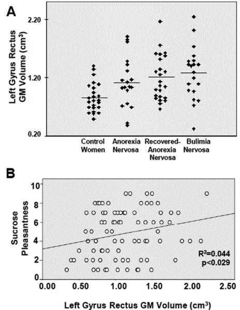
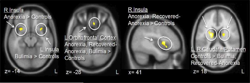
All eating disorder groups exhibited increased gray matter volume of the medial orbitofrontal cortex (gyrus rectus). Manual tracing confirmed larger gyrus rectus volume, and volume predicted taste pleasantness ratings across all groups. Analyses also indicated other morphological differences between diagnostic categories. Antero-ventral insula gray matter volumes were increased on the right side in the anorexia nervosa and recovered anorexia nervosa groups and on the left side in the bulimia nervosa group relative to the healthy comparison group. Dorsal striatum volumes were reduced in the recovered anorexia nervosa and bulimia nervosa groups and predicted sensitivity to reward in all three eating disorder groups. The eating disorder groups also showed reduced white matter in right temporal and parietal areas relative to the healthy comparison group. The results held when a range of covariates, such as age, depression, anxiety, and medications, were controlled for.
Brain structure in the medial orbitofrontal cortex, insula, and striatum is altered in eating disorders and suggests altered brain circuitry that has been associated with taste pleasantness and reward value.
神经性厌食症的病理生理学仍然不清楚,但结构脑改变可能是功能上重要的生物标志物。作者评估了味觉愉悦度和奖赏敏感性与大脑结构的关系,这可能与厌食症中常见的食物回避有关。
作者使用结构磁共振成像研究了当前限制型神经性厌食症(N=19)、从限制型神经性厌食症中恢复的女性(N=24)、神经性贪食症女性(N=19)和健康对照组女性(N=24)的灰质和白质体积。
所有饮食失调组都表现出内侧眶额皮质(rectus 回)灰质体积增加。手动追踪证实了更大的 rectus 回体积,并且体积预测了所有组的味觉愉悦度评分。分析还表明了不同诊断类别的其他形态差异。在神经性厌食症和恢复的神经性厌食症组中,右侧前腹侧岛叶灰质体积增加,在神经性贪食症组中左侧岛叶灰质体积增加,与健康对照组相比。背侧纹状体体积在恢复的神经性厌食症和神经性贪食症组中减少,并且预测了所有三组饮食失调症患者对奖赏的敏感性。与健康对照组相比,饮食失调症组还显示右侧颞叶和顶叶区域的白质减少。当控制了一系列协变量,如年龄、抑郁、焦虑和药物时,结果仍然成立。
内侧眶额皮质、岛叶和纹状体的大脑结构在饮食失调症中发生改变,表明与味觉愉悦度和奖赏价值相关的大脑回路改变。