Kim S, Lin L, Brown G A J, Hosaka K, Scott E W
Program in Stem Cell Biology and Regenerative Medicine, University of Florida College of Medicine, Gainesville, FL, USA.
Department of Molecular Genetics and Microbiology, University of Florida, Gainesville, FL, USA.
Leukemia. 2017 Jul;31(7):1582-1592. doi: 10.1038/leu.2016.354. Epub 2016 Nov 28.
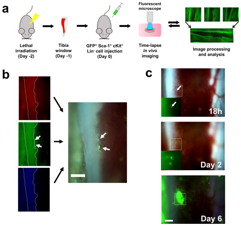
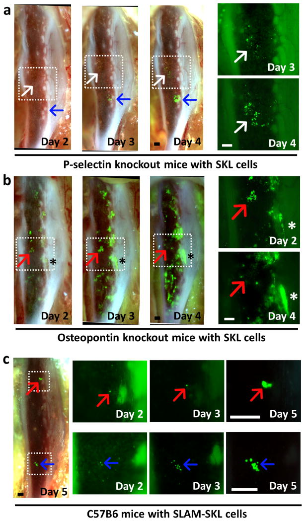
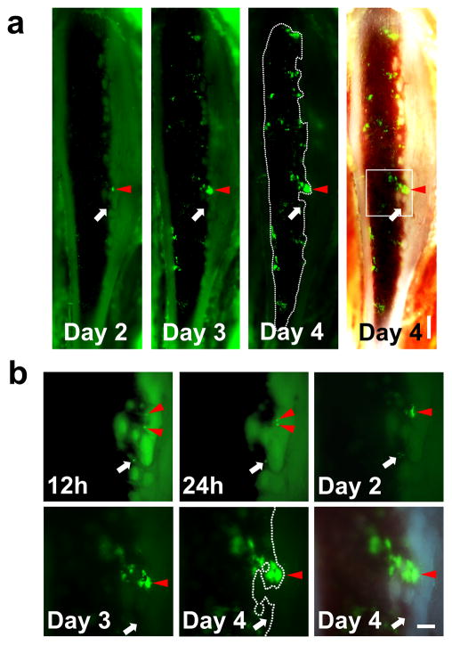
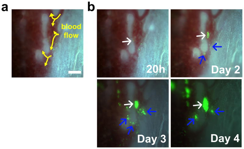
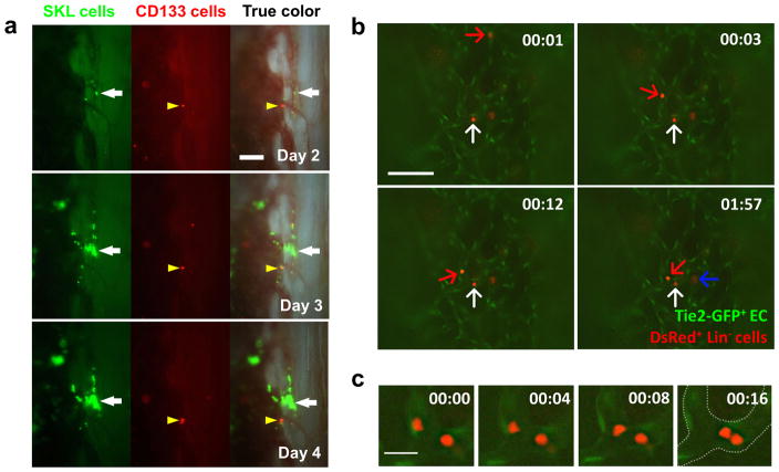
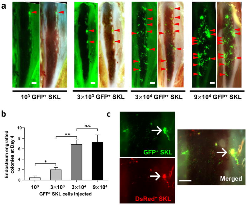
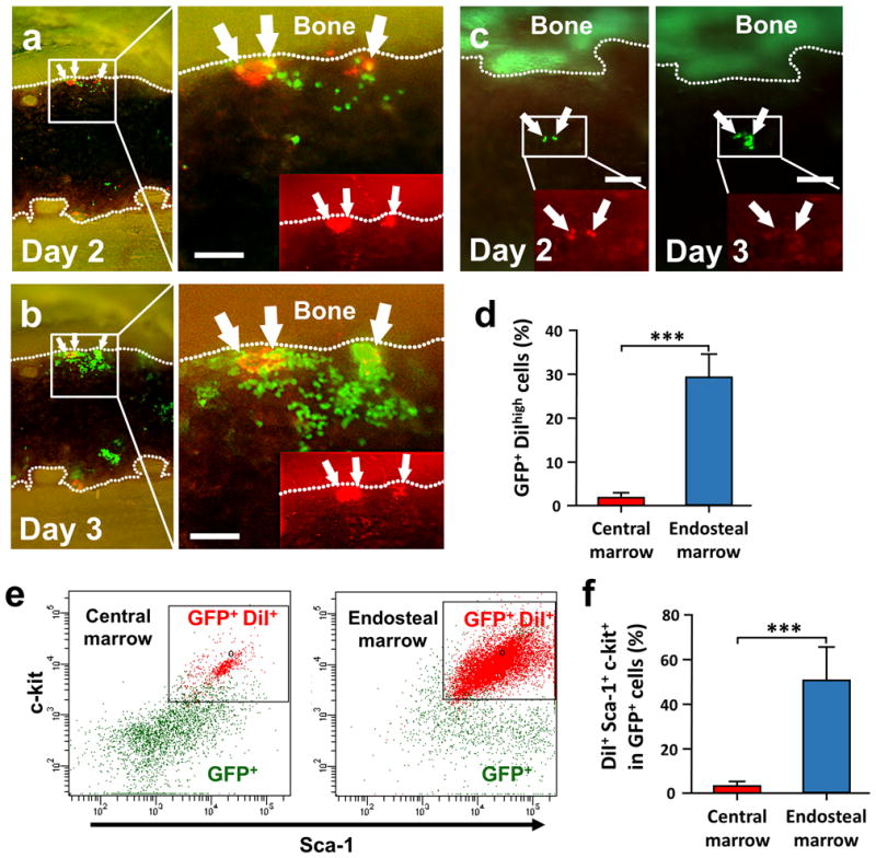
Homing, engraftment and proliferation of hematopoietic stem/progenitor cell (HSC/HPCs) are crucial steps required for success of a bone marrow transplant. Observation of these critical events is limited by the opaque nature of bone. Here we demonstrate how individual HSCs engraft in long bones by thinning one side of the tibia for direct and unbiased observation. Intravital imaging enabled detailed visualization of single Sca-1, c-Kit, Lineage (SKL) cell migration to bone marrow niches and subsequent proliferation to reconstitute hematopoiesis. This longitudinal study allowed direct observation of dynamic HSC/HPC activities during engraftment in full color for up to 6 days in live recipients. Individual SKL cells, but not mature or committed progenitor cells, preferentially homed to a limited number of niches near highly vascularized endosteal regions, and clonally expanded. Engraftment of SKL cells in P-selectin and osteopontin knockout mice showed abnormal homing and expansion of SKL cells. CD150, CD48 SKL populations initially engrafted in the central marrow region, utilizing only a subset of niches occupied by the parent SKL cells. Our study demonstrates that time-lapse imaging of tibia can be a valuable tool to understand the dynamic characteristics of functional HSC and niche components in various mouse models.
造血干/祖细胞(HSC/HPCs)的归巢、植入和增殖是骨髓移植成功所需的关键步骤。由于骨骼的不透明性,对这些关键事件的观察受到限制。在此,我们展示了如何通过将胫骨一侧变薄以进行直接且无偏差的观察,来观察单个造血干细胞在长骨中的植入情况。活体成像能够详细观察单个Sca-1、c-Kit、谱系(SKL)细胞向骨髓龛的迁移以及随后的增殖以重建造血功能。这项纵向研究能够在活体受体中对植入过程中造血干/祖细胞的动态活动进行长达6天的全彩色直接观察。单个SKL细胞,而非成熟或定向祖细胞,优先归巢至高度血管化的骨内膜区域附近数量有限的龛,并进行克隆性扩增。SKL细胞在P-选择素和骨桥蛋白基因敲除小鼠中的植入显示出SKL细胞归巢和扩增异常。CD150、CD48 SKL群体最初植入中央骨髓区域,仅利用亲代SKL细胞占据的一部分龛。我们的研究表明,对胫骨进行延时成像可成为了解各种小鼠模型中功能性造血干细胞和龛成分动态特征的宝贵工具。